. 2008 Mar;178(3):1237-49.
doi: 10.1534/genetics.107.083535. Epub 2008 Feb 3.

Recruitment and dissociation of nonhomologous end joining proteins at a DNA double-strand break in Saccharomyces cerevisiae

Affiliations

Recruitment and dissociation of nonhomologous end joining proteins at a DNA double-strand break in Saccharomyces cerevisiae

Dongliang Wu et al. Genetics. 2008 Mar.

Abstract

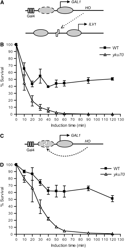
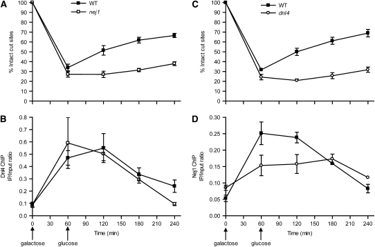
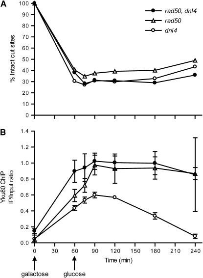
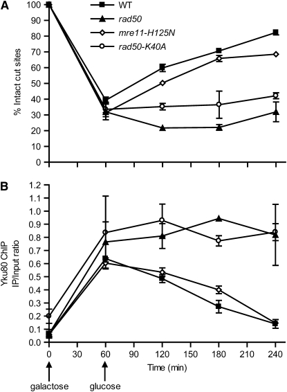
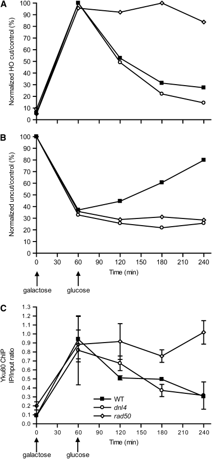
Nonhomologous end joining (NHEJ) is an important DNA double-strand-break (DSB) repair pathway that requires three protein complexes in Saccharomyces cerevisiae: the Ku heterodimer (Yku70-Yku80), MRX (Mre11-Rad50-Xrs2), and DNA ligase IV (Dnl4-Lif1), as well as the ligase-associated protein Nej1. Here we use chromatin immunoprecipitation from yeast to dissect the recruitment and release of these protein complexes at HO-endonuclease-induced DSBs undergoing productive NHEJ. Results revealed that Ku and MRX assembled at a DSB independently and rapidly after DSB formation. Ligase IV appeared at the DSB later than Ku and MRX and in a strongly Ku-dependent manner. Ligase binding was extensive but slightly delayed in rad50 yeast. Ligase IV binding occurred independently of Nej1, but instead promoted loading of Nej1. Interestingly, dissociation of Ku and ligase from unrepaired DSBs depended on the presence of an intact MRX complex and ATP binding by Rad50, suggesting a possible role of MRX in terminating a NHEJ repair phase. This activity correlated with extended DSB resection, but limited degradation of DSB ends occurred even in MRX mutants with persistently bound Ku. These findings reveal the in vivo assembly of the NHEJ repair complex and shed light on the mechanisms controlling DSB repair pathway utilization.


Figures

F<sc>igure</sc> 1.—
Figure 1.—
DSB systems used in this study. (A) DSB induction paradigm for ILV1-cs strains. Ovals represent nucleosomes, arrows represent transcription start sites, rectangles represent Gal4-binding sites, and zigzags represent the HO recognition site. The dashed nucleosome leaves the GAL1 promoter upon activation by galactose (Boeger et al. 2003; Reinke and Horz 2003). See text for details. (B) Survival assay of wild-type and yku70 mutant ILV1-cs strains. Yeast cells grown in galactose liquid medium for the indicated times were plated on glucose medium and incubated at 30° for 3 days. Colony counts at each time point are expressed as a percentage of the value at time 0. Data points are the mean ± SEM of three independent experiments. (C) DSB induction paradigm for GAL1-cs strains, similar to A. (D) Survival assay of GAL1-cs strains, similar to B. Time-dependent killing and repair in wild type are evident in both systems, but with slower cutting and more extensive repair with the GAL1-cs.
F<sc>igure</sc> 2.—
Figure 2.—
Appearance and disappearance of Yku80, Xrs2, and Dnl4 at a DSB. A DSB was induced in the GAL1 promoter (see Figure 1C) in various yeast strains by a 60-min galactose exposure followed by transfer to glucose to terminate HO expression. (A, C, and E) Chromatin fractions were prepared at different time points and portions used in the CQ–PCR assay to follow DSB formation and subsequent repair. (B, D, and F) Separate portions of the chromatin preparations were subjected to ChIP analysis using antibodies directed against tagged Yku80 (B), Xrs2 (D), and Dnl4 (F). Specific binding of each protein to the DSB is expressed as the immunoprecipitation-to-input PCR ratio as described in materials and methods. The complete experiment was performed at least two times for each strain with similar results; one representative experiment is shown. The mean ± SEM of two separate CQ–PCR reactions from the input DNA (A, C, and E) or ChIP samples (B, D, and F) of that experiment are shown.
F<sc>igure</sc> 3.—
Figure 3.—
Time course of initial binding of Yku80, Xrs2, and Dnl4 at a DSB. A DSB was induced in the ILV1 promoter (see Figure 1A) in a single yeast strain in which each of Yku80, Xrs2, and Lif1 were tagged for ChIP analysis with different antibodies. HO induction was initiated with galactose at time 0 and maintained throughout the experiment. (A) DSB monitoring similar to Figure 2, A, C, and E. (B) ChIP assay of Yku80, Xrs2, and Dnl4 similar to Figure 2, B, D, and F, performed on fractions of the same chromatin preparation as in A. (C) The same data in A and B normalized to allow plotting on the same graph. DSB formation is now expressed as the percentage of alleles broken, where 100% is the maximum level of breakage observed during the experiment. ChIP data were also normalized to span from the smallest to the largest values. Dnl4 binding was delayed ∼10 min relative to Yku80 and Xrs2 binding, which themselves could not be distinguished and occurred coincidentally with DSB formation at the earliest time points.
F<sc>igure</sc> 4.—
Figure 4.—
Dnl4 helps recruit Nej1 to a DSB, but not vice versa. (A and C) DSB monitoring in wild-type, nej1, and dnl4 strains similar to Figure 2, A, C, and E. (B and D) Dnl4 and Nej1 ChIP analysis, respectively, similar to Figure 2, B, D, and F, performed on fractions of the same chromatin preparations as in A and C.
F<sc>igure</sc> 5.—
Figure 5.—
Yku80 ChIP in rad50 dnl4 double-mutant yeast. (A) DSB monitoring in dnl4, rad50, and double-mutant strains, similar to Figure 2, A, C, and E. As expected, all strains are repair deficient. (B) Yku80 ChIP was performed in these same strains, similar to Figure 2B. rad50 mutation caused persistent binding of Yku80 even in the dnl4 background, demonstrating that this effect is specific to MRX function and not NHEJ efficiency.
F<sc>igure</sc> 6.—
Figure 6.—
MRX ATP-binding activity, but not nuclease activity, is required for Ku dissociation from a DSB. (A) DSB monitoring in wild-type, mre11-H125N, and rad50-K40A yeast, similar to Figure 2, A, C, and E. (B) ChIP assay of Yku80 in parallel with A, similar to Figure 2, B, D, and F.
F<sc>igure</sc> 7.—
Figure 7.—
Persistent Ku binding is correlated with reduced end resection. (A) DNAs prepared from wild type, rad50, dnl4, and yku70 yeast using the same time course as in Figure 2 were digested with HindIII and subjected to Southern blot analysis with probes specific to the GAL-cs and APN1 control. Uncut and HO-cut GAL-cs bands are indicated. Bands were subsequently quantified with a phosphorimager. (B) The ratio of the HO-cut band to the APN1 control was normalized to the ratio at 60 min, the time of HO shut off when DSB formation was maximal. Disappearance of the HO-cut band at subsequent times results from NHEJ (wild-type strain only) and/or resection (all strains). (C) The ratio of uncut HO band to the APN1 control was normalized to the ratio at time 0 to allow monitoring of DSB formation and repair in a manner analogous to the CQ–PCR method in previous figures.
F<sc>igure</sc> 8.—
Figure 8.—
Yku80 ChIP and Southern analysis of resection in G1 cells. (A and B) DSB analysis of resection and repair, similar to Figure 7, B and C, of wild-type, dnl4, and rad50 strains arrested in G1 by α-factor. (C) Yku80 ChIP, performed as in Figure 2, B, D, and F, except using cells maintained in G1 by α-factor arrest. MRX-dependent resection and Ku dissociation occurred in G1, but to a lesser final extent than in asynchronous cells.
F<sc>igure</sc> 9.—
Figure 9.—
LM–PCR analysis of resection reveals MRX-independent nucleotide loss. Wild-type, rad50, dnl4, and yku70 cells growing either exponentially (A) or arrested by α-factor (B) were induced to express HO with galactose and repressed with glucose 60 min later. LM–PCR was performed at various times as described in materials and methods using an oligonucleotide adapter capable of being ligated to an unresected HO-cut DSB. The signal from the LM–PCR product was expressed as a ratio relative to the AmpR control product and then normalized to the maximum value for each strain. LM–PCR signal persisted for a time and then disappeared in a manner independent of cell cycle stage, NHEJ, and MRX status.

Similar articles

Cited by

References

    1. Ahnesorg, P., and S. P. Jackson, 2007. The non-homologous end-joining protein Nej1p is a target of the DNA damage checkpoint. DNA Repair 6 190–201. - PMC - PubMed
    1. Anderson, D. E., K. M. Trujillo, P. Sung and H. P. Erickson, 2001. Structure of the Rad50 × Mre11 DNA repair complex from Saccharomyces cerevisiae by electron microscopy. J. Biol. Chem. 276 37027–37033. - PubMed
    1. Aparicio, O., J. V. Geisberg, E. Sekinger, A. Yang, Z. Moqtaderi et al., 2005. Chromatin immunoprecipitation for determining the association of proteins with specific genomic sequences in vivo, pp. 21.3.1–21.3.33 in Current Protocols in Molecular Biology, edited by F. M. Ausubel, R. Brent, R. E. Kingston, D. D. Moore, J. G. Seidman, J. A. Smith and K. Struhl. John Wiley & Sons, New York. - PubMed
    1. Aylon, Y., B. Liefshitz and M. Kupiec, 2004. The CDK regulates repair of double-strand breaks by homologous recombination during the cell cycle. EMBO J. 23 4868–4875. - PMC - PubMed
    1. Boeger, H., J. Griesenbeck, J. S. Strattan and R. D. Kornberg, 2003. Nucleosomes unfold completely at a transcriptionally active promoter. Mol. Cell 11 1587–1598. - PubMed

Publication types

MeSH terms