

doi: 10.1038/nn2047.
Epub 2008 Feb 3.
Astrocytes as determinants of disease progression in inherited amyotrophic lateral sclerosis
Affiliations
- PMID: 18246065
- PMCID: PMC3137510
- DOI: 10.1038/nn2047
Item in Clipboard
Astrocytes as determinants of disease progression in inherited amyotrophic lateral sclerosis
Nat Neurosci.
2008 Mar.
Abstract
Dominant mutations in superoxide dismutase cause amyotrophic lateral sclerosis (ALS), an adult-onset neurodegenerative disease that is characterized by the loss of motor neurons. Using mice carrying a deletable mutant gene, diminished mutant expression in astrocytes did not affect onset, but delayed microglial activation and sharply slowed later disease progression. These findings demonstrate that mutant astrocytes are viable targets for therapies for slowing the progression of non-cell autonomous killing of motor neurons in ALS.
Figures
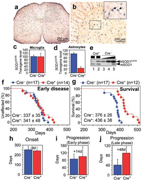
(a,b) β-galactosidase (β–gal) activity within astrocytes in (a) whole or (b) anterior horn region of the lumbar spinal cord section of GFAP-Cre/Rosa26 reporter mice visualized with X-gal and immunostaining with GFAP antibody. (Inset) Magnified image of the boxed area in (b). Arrows indicate β–Gal/GFAP-Cre expressing astrocytes. (c, d) LoxSOD1G37R transgene levels (n=3 for each group) in (c) primary microglia or (d) astrocytes from LoxSOD1G37R/GFAP-Cre+ and LoxSOD1G37R mice using real-time PCR. (e) SOD1G37R and mouse SOD1 content determined by immunoblotting in extracts from isolated primary astrocytes of (lanes 1, 2) LoxSOD1G37R/GFAP-Cre+ and (lanes 3–5) a dilution series of a comparable extract from LoxSOD1G37R astrocytes representing 25%, 50% and 100% of the protein amounts loaded in lanes 1 and 2. Ages at which (f) early disease phase (to 10% weight loss) (p=0.76) or (g) end-stage (p<0.0001) was reached for (red) LoxSOD1G37R/GFAP-Cre+ mice and (blue) LoxSOD1G37R littermates. Mean ages ± standard deviation were provided. (h–j) Mean onset (p=0.47) (h), mean duration of early disease (p=0.35) (i) (from onset to 10% weight loss) and a later disease (p<0.0001) (j) (from 10% weight loss to end-stage) for (red) LoxSOD1G37R/GFAP-Cre+ and (blue) LoxSOD1G37R littermates. At each time point, p value was determined by unpaired t test. Error bars denote standard deviation.
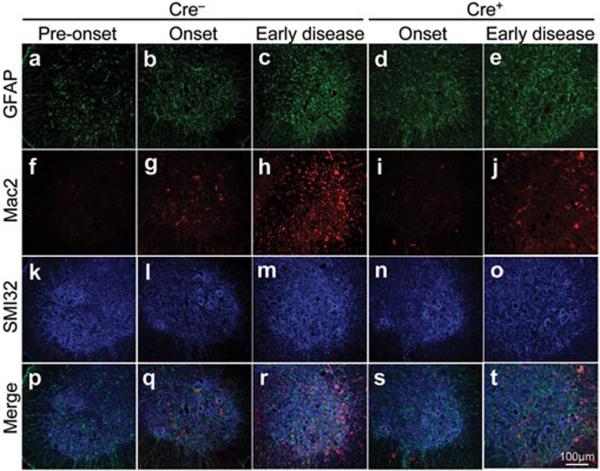
(a–e) GFAP detected within astrocytes, (f–j) Mac2-positive activated microglia, and (k–o) motor neurons identified with the neurofilament antibody SMI-32 staining in the lumbar spinal cord of a LoxSOD1G37R mouse (a, f, k, p) prior to disease onset, (b, g, l, p) at disease onset, or (c, h, m, r) at early disease, together with LoxSOD1G37R/GFAP-Cre+ mice (d, i, n, s) at onset or (e, j, o, t) early disease. (p–t) Merged images.
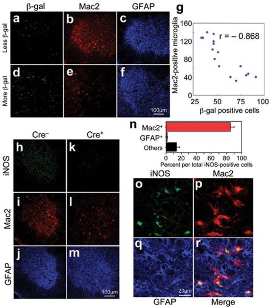
(a–f) Images of (a, d) β-galactosidase, (b, e) Mac2, and (c, f) GFAP staining from a (a–c) left and (d–f) right lumbar spinal cord section from a 12-month-old LoxSOD1G37R/GFAP-Cre+ mouse. (a, d) GFAP-Cre+ astrocytes are marked by β-galactosidase. (g) Inverted correlation between the number of Cre-positive astrocytes and Mac2-positive microglia in LoxSOD1G37R/GFAP-Cre+ mice lumbar spinal cord sections. Correlation coefficient r= −0.868 (p<0.001). (h–m) Lumbar spinal cord sections from (h–j) LoxSOD1G37R and (k–m) LoxSOD1G37R/GFAP-Cre+ mice at early disease stage immunostained with antibodies to (h, k) iNOS, (i, l) Mac2, and (j, m) GFAP. (n) Quantification of iNOS-positive cells within anterior horn from lumbar spinal cord of symptomatic LoxSOD1G37R mice. Averaged percent of iNOS+/Mac2+ (red), iNOS+/GFAP+ (blue), and iNOS+/other cell type (black) per total iNOS+ cells was plotted. (o–r) Magnified images of anterior horn from lumbar spinal cord of symptomatic LoxSOD1G37R mice stained with (o) iNOS, (p) Mac2, and (q) GFAP. (r) Merged image illustrates that iNOS positive cells are Mac2-positive microglia.
References
Publication types
MeSH terms
Substances
Grants and funding
LinkOut - more resources
Full Text Sources
Other Literature Sources
Medical
Molecular Biology Databases
Miscellaneous
