. 2008 Feb;14(2):125-33.
doi: 10.1038/nm1725. Epub 2008 Feb 3.

Prevention of the neurocristopathy Treacher Collins syndrome through inhibition of p53 function

Affiliations

Prevention of the neurocristopathy Treacher Collins syndrome through inhibition of p53 function

Natalie C Jones et al. Nat Med. 2008 Feb.

Abstract

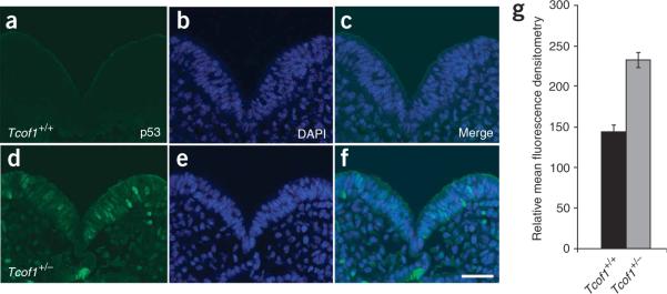
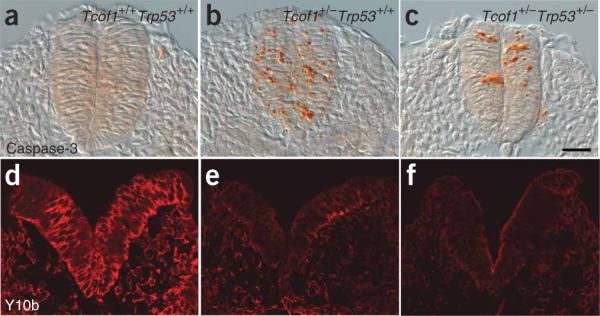
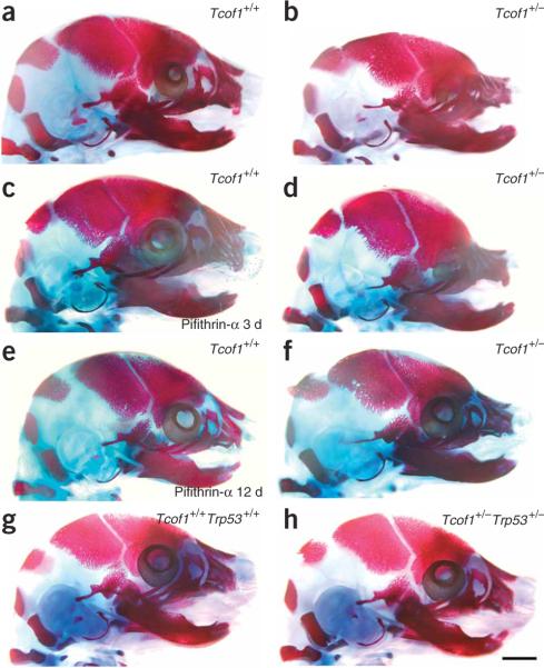
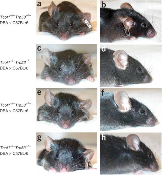
Treacher Collins syndrome (TCS) is a congenital disorder of craniofacial development arising from mutations in TCOF1, which encodes the nucleolar phosphoprotein Treacle. Haploinsufficiency of Tcof1 perturbs mature ribosome biogenesis, resulting in stabilization of p53 and the cyclin G1-mediated cell-cycle arrest that underpins the specificity of neuroepithelial apoptosis and neural crest cell hypoplasia characteristic of TCS. Here we show that inhibition of p53 prevents cyclin G1-driven apoptotic elimination of neural crest cells while rescuing the craniofacial abnormalities associated with mutations in Tcof1 and extending life span. These improvements, however, occur independently of the effects on ribosome biogenesis; thus suggesting that it is p53-dependent neuroepithelial apoptosis that is the primary mechanism underlying the pathogenesis of TCS. Our work further implies that neuroepithelial and neural crest cells are particularly sensitive to cellular stress during embryogenesis and that suppression of p53 function provides an attractive avenue for possible clinical prevention of TCS craniofacial birth defects and possibly those of other neurocristopathies.


Figures

Figure 1
Figure 1
Immunoblot analysis of p53. (a) Analysis of the 53-kDa protein in E10.5 Tcof1+/+ embryos (lanes 1 and 2) as compared to E10.5 Tcof1+/− littermates (lanes 3 and 4). α-tubulin was used as a protein loading control. (b) Bar graph denistometry quantification of p53 protein levels in Tcof1+/+ and mutant Tcof1+/− littermates. Statistical significance was determined with the one-tailed Student's t-test with P = 0.006.
Figure 2
Figure 2
Immunostaining of p53. Immunostaining of E8.5 embryos for p53 protein (green) in Tcof1+/+ embryos (ac) and inTcof1+/− embryos (df). Counterstaining with DAPI (blue; b,e) shows that p53 is specifically stabilized in the nucleus of neuroepithelial cells in Tcof1+/− embryos (f). Scale bar, 40 mm. (g) Bar graph of mean fluorescence denistometry quantification of p53 protein levels in individual neuroepithelial cells of Tcof1+/+ and Tcof1+/− littermates. Statistical significance was determined with the one-tailed Student's t-test with P < 0.001.
Figure 3
Figure 3
Expression of Ccng1 and inhibition of apoptosis. (ad) In situ hybridization for Ccng1 in whole (a,b) and sectioned (c,d) E8.5 embryos. (e,f) TUNEL staining (green) for apoptosis in E8.75 Tcof1+/+ and Tcof1+/− littermates. (g,h) Intraperitoneal injection of pregnant dams with pifithrin-α in wild-type and Tcof1+/− embryos. Scale bars, 150 μm (a,b,eg); 40 μm (c,d).
Figure 4
Figure 4
Analyses of apoptosis and ribosome biogenesis. (ac) Immunostaining of sections of E9.0–E9.5 embryos for caspase3 activity (orange) in wild-type Tcof1+/+ embryos (a), Tcof1+/− mutants (b) and in Tcof1+/−Trp53+/− littermates (c). (df) Immunostaining of sections of E8.5 embryos with the mature 28S rRNA-specific antibody Y10b (red) in Tcof1+/+ embryos (d), Tcof1+/− mutants (e) and in Tcof1+/− Trp53+/− embryo littermates (f). Scale bar, 50 μm.
Figure 5
Figure 5
Embryonic skeletal analyses and prevention of TCS. Craniofacial skeletons of E17.5 embryos were stained for bone (alizarin red) and cartilage (alcian blue). (a) Wild-type Tcof1+/+ embryo, showing a normal craniofacial skeleton. (b) Mutant Tcof1+/− embryo, showing severe frontonasal hypoplasia including the nasal, frontal, premaxilla, maxilla and mandibular bones, and doming of the skull and severe microphthalmia. (c) Tcof1+/+ embryo treated with pifithrin-α for 3 d in utero, showing a normal craniofacial skeleton. (d) Tcof1+/− embryo treated with pifithrin-α for 3 d in utero, showing slightly reduced frontonasal hypoplasia and less severe microphthalmia. (e) Tcof1+/+ embryo treated with pifithrin-α for 12 d in utero, showing a normal craniofacial skeleton. (f) Tcof1+/− embryo treated with pifithrin-α for 12 d in utero, showing substantially reduced frontonasal hypoplasia with considerable restoration of normal patterning of the frontal, nasal, premaxilla, maxilla and mandibular bones, and little evidence of any persistent calvarial doming or microphthalmia. (g) Wild-type Tcof1+/+Trp53+/+ embryo, showing a normal craniofacial skeleton. (h) Tcof1+/−Trp53+/− littermate embryo, showing a completely normal craniofacial skeleton that is indistinguishable from wild-type. Scale bar, 2 mm.
Figure 6
Figure 6
Genetic prevention of postnatal TCS craniofacial anomalies. Shown are frontal and lateral views highlighting restoration of normal craniofacial morphology through loss of p53 function. (a,b) Tcof1+/+Trp53+/− mouse. (c,d) Tcof1+/+Trp53−/− mouse. (e,f) Tcof1+/−Trp53+/− mouse. (g,h) Tcof1+/−Trp53−/− mouse. All mice were on a mixed DBA × C57BL/6 background.

Comment in

References

    1. Phelps PD, Poswillo D, Lloyd GA. The ear deformities in mandibulofacial dysostosis (Treacher Collins syndrome) Clin. Otolaryngol. 1981;6:15–28. - PubMed
    1. Rovin S, Dachi SF, Borenstein DB, Cotter WB. Mandibulofacial dysostosis, a familial study of five generations. J. Pediatr. 1964;65:215–221. - PubMed
    1. Fazen LE, Elmore J, Nadler HL. Mandibulo-facial dysostosis. (Treacher-Collins syndrome) Am. J. Dis. Child. 1967;113:405–410. - PubMed
    1. Edwards SJ, Gladwin AJ, Dixon MJ. The mutational spectrum in Treacher Collins syndrome reveals a predominance of mutations that create a premature-termination codon. Am. J. Hum. Genet. 1997;60:515–524. - PMC - PubMed
    1. Gladwin AJ, et al. Treacher Collins syndrome may result from insertions, deletions or splicing mutations, which introduce a termination codon into the gene. Hum. Mol. Genet. 1996;5:1533–1538. - PubMed

Publication types

MeSH terms

Associated data