

Review
doi: 10.1172/JCI34006.
Stressed out: the skeletal muscle ryanodine receptor as a target of stress
Affiliations
- PMID: 18246195
- PMCID: PMC2214709
- DOI: 10.1172/JCI34006
Item in Clipboard
Review
Stressed out: the skeletal muscle ryanodine receptor as a target of stress
J Clin Invest.
2008 Feb.
Abstract
Over the past century, understanding the mechanisms underlying muscle fatigue and weakness has been the focus of much investigation. However, the dominant theory in the field, that lactic acidosis causes muscle fatigue, is unlikely to tell the whole story. Recently, dysregulation of sarcoplasmic reticulum (SR) Ca(2+) release has been associated with impaired muscle function induced by a wide range of stressors, from dystrophy to heart failure to muscle fatigue. Here, we address current understandings of the altered regulation of SR Ca(2+) release during chronic stress, focusing on the role of the SR Ca(2+) release channel known as the type 1 ryanodine receptor.
Figures
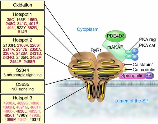
RyR1 forms a macromolecular complex with PDE4D3, A-kinase anchor protein (mAKAP), the catalytic (cat) and regulatory (reg) subunits of PKA, calstabin1, calmodulin, PP1, and spinophilin. Components of the RyR1 complex are represented schematically bound to one monomer of the homotetrameric RyR1 channel. On the left, stress signals targeting RyR1 are shown, including the three hotspots on RyR1 where disease-causing mutations cluster (34, 114). MH-associated mutations are in red, CCD-associated mutations are in pink, mixed mutations or those associated with multi-minicore disease or nemaline rod disease are shown in black. –, deletion mutation.
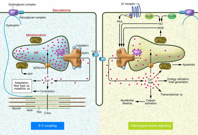
Depolarization of the T-tubule membrane activates Cav1.1, triggering SR Ca2+ release through RyR1 and leading to sarcomere contraction, a process known as E-C coupling. Intracellular signaling pathways activated in skeletal muscle by pathological stress affect RyR1 function and alter E-C coupling. Stress-induced RyR1 dysfunction can result in SR Ca2+ leak, which potentially activates numerous Ca2+-dependent cellular damage mechanisms. AC, adenylate cyclase.
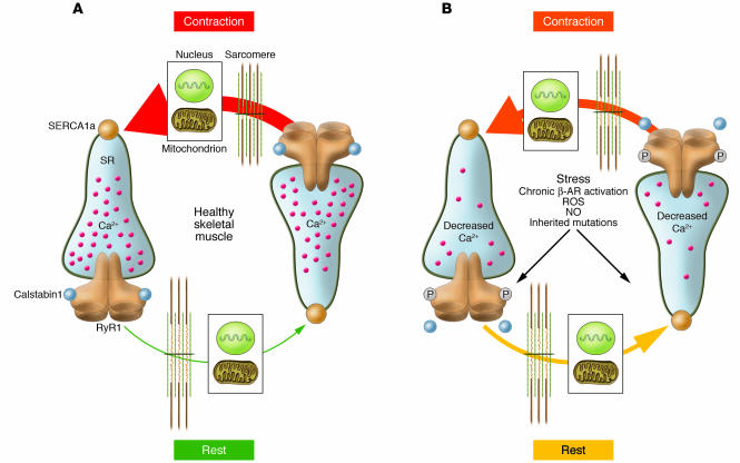
(A) In healthy muscle, Ca2+ release occurs in a coordinated fashion during contraction, and [Ca2+]cyt is low at rest. Organelles (e.g., the nucleus and mitochondria) sense changes in [Ca2+]cyt, which regulates cellular functions including transcription and energy metabolism. (B) Stress-induced PKA-mediated phosphorylation of RyR1 alters the way skeletal muscle handles Ca2+ during contraction and relaxation. PKA hyperphosphorylation of RyR1, resulting in calstabin1 depletion from the channel complex, leads to an SR Ca2+ leak in the resting muscle, potentially influencing nuclear and mitochondrial function. Ca2+ leak also decreases SR load, and less Ca2+ is available for release during contraction. β-AR, β-adrenergic receptor.
References
-
- Tanabe T., Beam K.G., Adams B.A., Niidome T., Numa S. Regions of the skeletal muscle dihydropyridine receptor critical for excitation-contraction coupling. Nature. 1990;346:567–569. - PubMed
-
- Fill M., Copello J.A. Ryanodine receptor calcium release channels. Physiol. Rev. 2002;82:893–922. - PubMed
-
- Gonzalez-Serratos H., Valle-Aguilera R., Lathrop D.A., Garcia M.C. Slow inward calcium currents have no obvious role in muscle excitation-contraction coupling. Nature. 1982;298:292–294. - PubMed
-
- Rios E., Brum G. Involvement of dihydropyridine receptors in excitation-contraction coupling in skeletal muscle. Nature. 1987;325:717–720. - PubMed
-
- Catterall W.A. Excitation-contraction coupling in vertebrate skeletal muscle: a tale of two calcium channels. Cell. 1991;64:871–874. - PubMed
Publication types
MeSH terms
Substances
LinkOut - more resources
Full Text Sources
Other Literature Sources
Medical
Research Materials
Miscellaneous
