

A new system for measuring three-dimensional back shape in scoliosis
- PMID: 18247064
- PMCID: PMC2367415
- DOI: 10.1007/s00586-007-0581-x
A new system for measuring three-dimensional back shape in scoliosis
Abstract
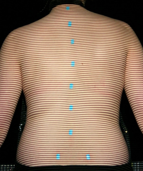
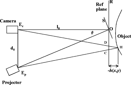
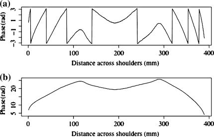
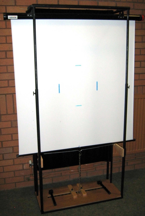
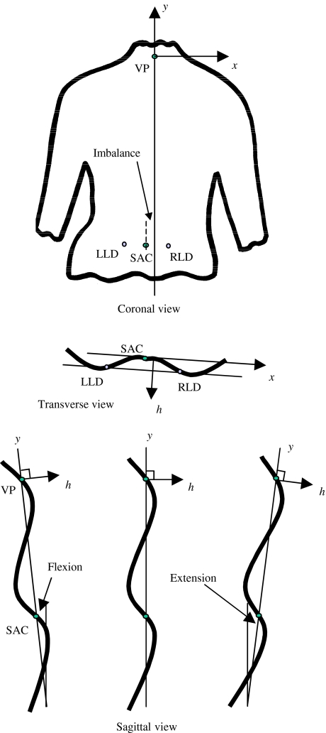
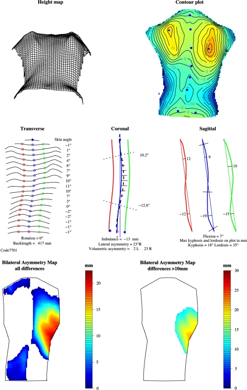
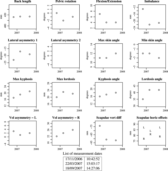
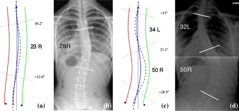
The aim of this work was to develop a low-cost automated system to measure the three-dimensional shape of the back in patients with scoliosis. The resulting system uses structured light to illuminate a patient's back from an angle while a digital photograph is taken. The height of the surface is calculated using Fourier transform profilometry with an accuracy of +/-1 mm. The surface is related to body axes using bony landmarks on the back that have been palpated and marked with small coloured stickers prior to photographing. Clinical parameters are calculated automatically and presented to the user on a monitor and as a printed report. All data are stored in a database. The database can be interrogated and successive measurements plotted for monitoring the deformity changes. The system developed uses inexpensive hardware and open source software. Accurate surface topography can help the clinician to measure spinal deformity at baseline and monitor changes over time. It can help the patients and their families to assess deformity. Above all it reduces the dependence on serial radiography and reduces radiation exposure when monitoring spinal deformity.
Figures
References
-
- Adair I, VanWijk M, Armstrong G. Moiré topography in scoliosis screening. Clin Orthop Relat Res. 1977;129:165–171. - PubMed
-
- Berryman F (2004) Fourier transform profilometry for measuring back shape in scoliosis. PhD. School of Engineering and the Built Environment, University of Wolverhampton, Wolverhampton
-
- Berryman F, Gardner A, Pynsent P, Fairbank J (2007) The relationship between Cobb angle and ISIS2 lateral asymmetry. In: Scoliosis Research Society eastern european regional meeting. Budapest, Hungary
-
- Berryman F, Pynsent P, Cubillo J. The effect of windowing in Fourier transform profilometry applied to noisy images. Opt Lasers Eng. 2004;41(6):815–825. doi: 10.1016/S0143-8166(03)00061-7. - DOI
Publication types
MeSH terms
LinkOut - more resources
Full Text Sources
Other Literature Sources
Medical
