Merging molecular imaging and RNA interference: early experience in live animals
- PMID: 18247325
- PMCID: PMC4836383
- DOI: 10.1002/jcb.21689
Merging molecular imaging and RNA interference: early experience in live animals
Abstract
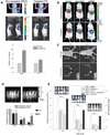
The rapid development of non-invasive imaging techniques and imaging reporters coincided with the enthusiastic response that the introduction of RNA interference (RNAi) techniques created in the research community. Imaging in experimental animals provides quantitative or semi-quantitative information regarding the biodistribution of small interfering RNAs and the levels of gene interference (i.e., knockdown of the target mRNA) in living animals. In this review we give a brief summary of the first imaging findings that have potential for accelerating the development and testing of new approaches that explore RNAi as a method for achieving loss-of-function effects in vivo and as a promising therapeutic tool.
2008 Wiley-Liss, Inc.
Figures


Similar articles
-
[Research progress of radionuclide tracing in small interference RNA imaging in vivo].Beijing Da Xue Xue Bao Yi Xue Ban. 2010 Oct 18;42(5):616-8, 1 p following 618. Beijing Da Xue Xue Bao Yi Xue Ban. 2010. PMID: 20957026 Review. Chinese.
-
In vivo imaging of oligonucleotide delivery.Methods Mol Biol. 2012;872:243-53. doi: 10.1007/978-1-61779-797-2_17. Methods Mol Biol. 2012. PMID: 22700416
-
Imaging of siRNA delivery and silencing.Methods Mol Biol. 2009;487:93-110. doi: 10.1007/978-1-60327-547-7_5. Methods Mol Biol. 2009. PMID: 19301644 Review.
-
RNA interference--a silent but an efficient therapeutic tool.Appl Biochem Biotechnol. 2013 Mar;169(6):1774-89. doi: 10.1007/s12010-013-0098-1. Epub 2013 Jan 23. Appl Biochem Biotechnol. 2013. PMID: 23340870 Review.
-
Single-Cell Imaging Approaches for Studying Small-RNA-Induced Gene Regulation.Biophys J. 2018 Jul 17;115(2):203-208. doi: 10.1016/j.bpj.2018.05.040. Epub 2018 Jun 30. Biophys J. 2018. PMID: 29970232 Free PMC article. Review.
Cited by
-
Technologies for investigating the physiological barriers to efficient lipid nanoparticle-siRNA delivery.J Histochem Cytochem. 2013 Jun;61(6):407-20. doi: 10.1369/0022155413484152. Epub 2013 Mar 14. J Histochem Cytochem. 2013. PMID: 23504369 Free PMC article. Review.
-
Knocking down barriers: advances in siRNA delivery.Nat Rev Drug Discov. 2009 Feb;8(2):129-38. doi: 10.1038/nrd2742. Nat Rev Drug Discov. 2009. PMID: 19180106 Free PMC article. Review.
-
Fluorocarbons Enhance Intracellular Delivery of Short STAT3-sensors and Enable Specific Imaging.Theranostics. 2017 Aug 3;7(13):3354-3368. doi: 10.7150/thno.19704. eCollection 2017. Theranostics. 2017. PMID: 28900515 Free PMC article.
-
Imaging CXCR4 Expression with (99m)Tc-Radiolabeled Small-Interference RNA in Experimental Human Breast Cancer Xenografts.Mol Imaging Biol. 2016 Jun;18(3):353-9. doi: 10.1007/s11307-015-0899-4. Mol Imaging Biol. 2016. PMID: 26452556
-
In vivo imaging of RNA interference.J Nucl Med. 2010 Feb;51(2):169-72. doi: 10.2967/jnumed.109.066878. Epub 2010 Jan 15. J Nucl Med. 2010. PMID: 20080892 Free PMC article. Review.
References
-
- Arwert E, Hingtgen S, Figueiredo JL, Bergquist H, Mahmood U, Weissleder R, Shah K. Visualizing the dynamics of EGFR activity and antiglioma therapies in vivo. Cancer Res. 2007;67:7335–7342. - PubMed
-
- Banan M, Puri N. The ins and outs of RNAi in mammalian cells. Curr Pharm Biotechnol. 2004;5:441–450. - PubMed
-
- Bartlett D, Davis M. Impact of tumor-specific targeting and dosing schedule on tumor growth inhibition after intravenous administration of siRNA-containing nanoparticles. Biotechnol Bioeng. 2007a 10.1002/bit.21668. - PubMed
-
- Bartlett DW, Davis ME. Effect of siRNA nuclease stability on the in vitro and in vivo kinetics of siRNA-mediated gene silencing. Biotechnol Bioeng. 2007b;97:909–921. - PubMed
Publication types
MeSH terms
Substances
Grants and funding
LinkOut - more resources
Full Text Sources
Other Literature Sources
Medical

