. 2008 Feb 8;4(2):e20.
doi: 10.1371/journal.ppat.0040020.

The macaque gut microbiome in health, lentiviral infection, and chronic enterocolitis

Affiliations

The macaque gut microbiome in health, lentiviral infection, and chronic enterocolitis

Philip McKenna et al. PLoS Pathog. .

Abstract

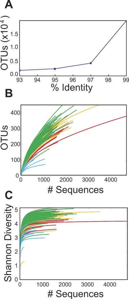
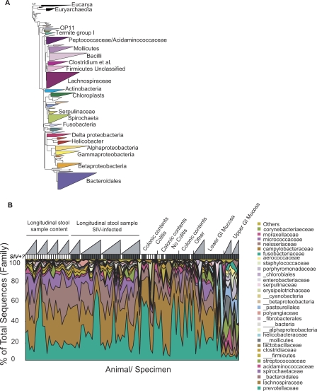
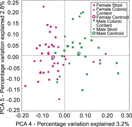
The vertebrate gut harbors a vast community of bacterial mutualists, the composition of which is modulated by the host immune system. Many gastrointestinal (GI) diseases are expected to be associated with disruptions of host-bacterial interactions, but relatively few comprehensive studies have been reported. We have used the rhesus macaque model to investigate forces shaping GI bacterial communities. We used DNA bar coding and pyrosequencing to characterize 141,000 sequences of 16S rRNA genes obtained from 100 uncultured GI bacterial samples, allowing quantitative analysis of community composition in health and disease. Microbial communities of macaques were distinct from those of mice and humans in both abundance and types of taxa present. The macaque communities differed among samples from intestinal mucosa, colonic contents, and stool, paralleling studies of humans. Communities also differed among animals, over time within individual animals, and between males and females. To investigate changes associated with disease, samples of colonic contents taken at necropsy were compared between healthy animals and animals with colitis and undergoing antibiotic therapy. Communities from diseased and healthy animals also differed significantly in composition. This work provides comprehensive data and improved methods for studying the role of commensal microbiota in macaque models of GI diseases and provides a model for the large-scale screening of the human gut microbiome.


Conflict of interest statement

Competing interests. The authors have declared that no competing interests exist.

Figures

Figure 1
Figure 1. Use of DNA Bar-coding and Pyrosequencing to Analyze Uncultured Bacterial Communities
(A) Length distribution of the pyrosequence reads used in this study. The median length was 264 nt. (B) The DNA bar-coding strategy. PCR primers are indicated by the arrows, DNA 5′ ends are shown as balls. Each primer contains a region complementary to the 454 sequencing primers (either A or B) and the 16S rRNA gene DNA (either BSR357 or BSF8) separated by a unique 4 base bar code (bold). (C) Reproducibility of the pyrosequencing method. DNA from a single specimen was analyzed by pyrosequencing at two different centers. Bacterial orders are indicated by the color code. (D) Comparison of results with pyrosequencing and conventional Sanger sequencing. Bacterial orders are indicated by the color code. Numbers of sequences are as follows: M3T1 pyrosequence, 1382; M3T1 Sanger sequence, 47; M3T6 pyrosequence, 1360; M3T6 Sanger sequence, 47.
Figure 2
Figure 2. Diversity of the Macaque GI Microbiota
(A) The numbers of operational taxonomic units (OTUs) present in the collection of pyrosequence reads was analyzed by condensing sequences at several percent identity thresholds. The x-axis shows the percent identity, the y-axis the number of OTUs detected. (B) Collectors curves analysis of the completeness of sampling. Repeated samples of OTU subsets were used to evaluate whether further sampling would likely yield additional taxa (rarefaction analysis), as indicated by whether the curve has reached a plateau value. The y-axis indicates the number of OTUs detected, the x-axis the number of taxa in the sequence subset analyzed. The color codes are as follows: green, stool samples; yellow, colonic contents; red, lower GI mucosal surface; blue, upper GI mucosal surface. (C) Rarefaction curves to estimate the diversity of taxa present in individual samples, using the Shannon Index. Color code as in (B). The upper GI mucosal samples were significantly less diverse than the other groups (p < 0.004 for pairwise comparisons of upper gut samples to each of the other three; Mann-Whitney comparison of means).
Figure 3
Figure 3. Comparison of the Macaque GI Microbiota to That of Mice and Humans
The plot was generated using unweighted UniFrac. Mouse and human sequences were trimmed to match the macaque pyrosequence reads in length (264 nt) and location within the 16S rRNA gene. The differences among communities from the different vertebrates was significant at p < 0.001 (t-test with permutation).
Figure 4
Figure 4. Bacteria Composing the Macaque GI Microbiome
(A) Bacterial taxa identified from pyrosequencing data after alignment with the ARB 16S rRNA gene database. The size of each triangle indicates the relative number of OTUs within each taxa (100% identify threshold). (B) Summary of the bacterial taxa present in each gut community sampled, indicating the individual and temporal variation in the macaque GI microbiota. Each sample analyzed is indicated along the x-axis, the y-axis indicates the percent of the community comprised by each type of bacteria. A key to the bacterial taxa is listed at the right. Taxa corresponding to bacterial phyla are indicated with the triple underscore before the name, classes by a double underscore, orders by single underscores, and families by no underscore. Specific values for each community, along with clinical parameters for each monkey studied are summarized in Table S1.
Figure 5
Figure 5. Distinctive GI Microbiota in Samples from Communities from Different Anatomical Sites
Unweighted UniFrac was used in the comparison. The types of samples studied are indicated by the key at lower right.
Figure 6
Figure 6. Sexual Dimorphism in the Macaque GI Microbiota
Samples of stool and colonic contents are combined for this analysis. Cluster analysis was carried out using unweighted UniFrac. Separation between male communities (green) and female communities (pink) was significant (p < 0.05, t-test with permutation; analysis over all variation between samples). Note that with the simplest null model, we expect each Principal Coordinate to explain 100/number of samples, which is 100/100 communities = 1% of the variation. Thus the fourth Principal Coordinate, which separates males and females, is expected to contain meaningful information.
Figure 7
Figure 7. Colitis Is Associated with Distinctive GI Microbiota in Samples of Colonic Contents Taken at Necropsy
The analysis was restricted to samples of colonic contents taken at necropsy that allowed unambiguous assignment to the “colitis” or “healthy” categories. (A) Analysis of communities in unweighted UniFrac. Samples in the colitis and healthy categories showed significant separation along the first principal coordinate (p < 0.05, t-test with permutation). For an additional four animals, insufficient clinical histories were available, so these were not included in the analysis (Table 1). (B) Diversity in samples from healthy animals or those with colitis were analyzed using the Shannon Index on OTUs condensed at 97% identity. The diversity in the samples from animals with colitis was significantly lower (p < 0.05; Mann-Whitney comparison of means).

Similar articles

Cited by

References

    1. Ley RE, Peterson DA, Gordon JI. Ecological and evolutionary forces shaping microbial diversity in the human intestine. Cell. 2006;124:837–484. - PubMed
    1. Whitman WB, Coleman DC, Wiebe WJ. Prokaryotes: the unseen majority. Proc Natl Acad Sci U S A. 1998;95:6578–6583. - PMC - PubMed
    1. Gill SR, Pop M, Deboy RT, Eckburg PB, Turnbaugh PJ, et al. Metagenomic analysis of the human distal gut microbiome. Science. 2006;312:1355–1359. - PMC - PubMed
    1. Eckburg PB, Bik EM, Bernstein CN, Purdom E, Dethlefsen L, et al. Diversity of the human intestinal microbial flora. Science. 2005;308:1635–1638. - PMC - PubMed
    1. Palmer C, Bik EM, Eisen MB, Eckburg PB, Sana TR, et al. Rapid quantitative profiling of complex microbial populations. Nucleic Acids Res. 2006;10:e5. - PMC - PubMed

Publication types

MeSH terms