. 2008 Sep;76(7):820-30.
doi: 10.1111/j.1432-0436.2007.00263.x. Epub 2008 Jan 31.

Inhibition of monoamine oxidase A promotes secretory differentiation in basal prostatic epithelial cells

Affiliations

Inhibition of monoamine oxidase A promotes secretory differentiation in basal prostatic epithelial cells

Hongjuan Zhao et al. Differentiation. 2008 Sep.

Abstract

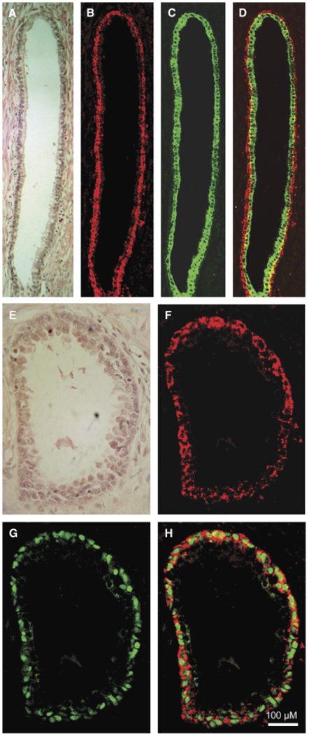
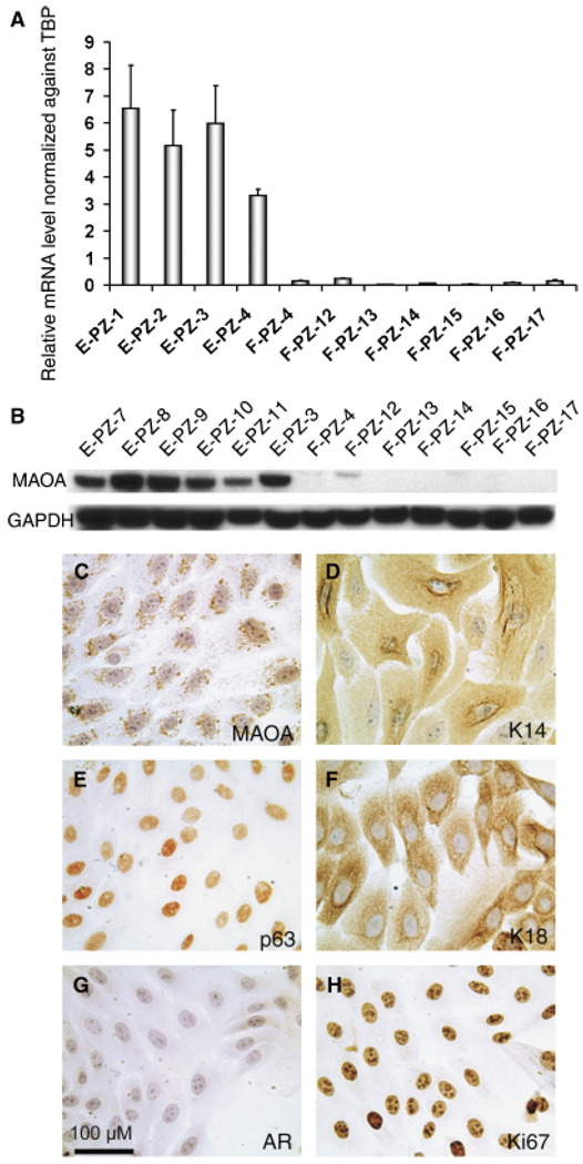
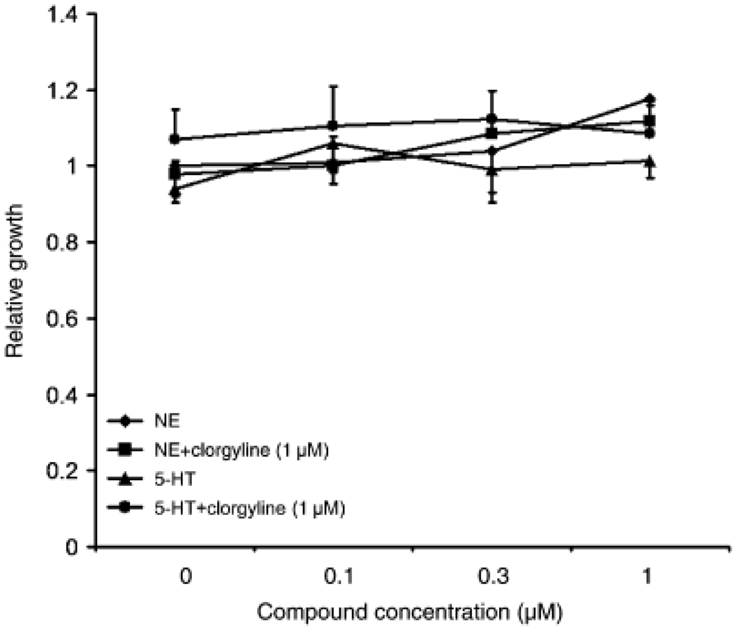
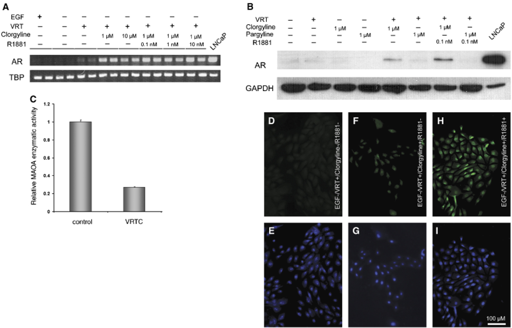
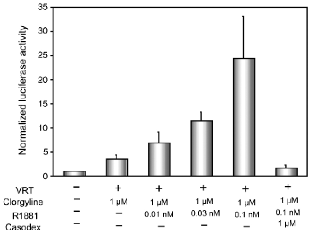
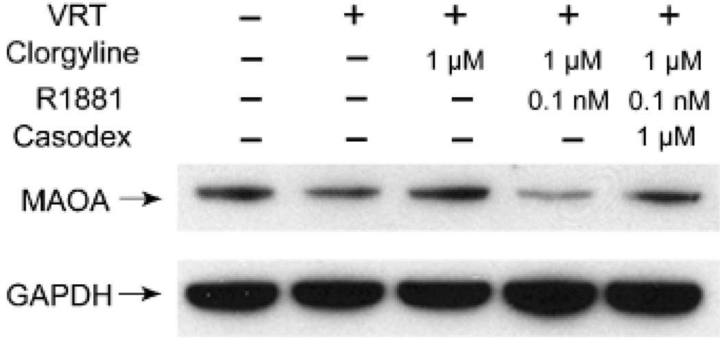
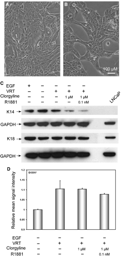
Monoamine oxidase A (MAO-A) expression is associated with high-grade prostate cancer. Immunohistochemistry showed that MAO-A is also expressed in the basal epithelial cells of normal prostate glands. Using cultured primary prostatic epithelial cells as a model, we showed that MAO-A prevents basal epithelial cells from differentiating into secretory cells. Under differentiation-promoting conditions, clorgyline, an irreversible MAO-A inhibitor, induced secretory cell-like morphology and repressed expression of cytokeratin 14, a basal cell marker. More importantly, clorgyline induced mRNA and protein expression of androgen receptor (AR), a hallmark of secretory epithelial cells. In clorgyline-treated cells, androgen induced luciferase activity controlled by the promoter of prostate-specific antigen, an AR target gene, in a dose-dependent manner. This activity was blocked by the AR antagonist Casodex, showing that AR is functional. In turn, androgen decreased MAO-A expression in clorgyline-treated, secretory-like cells. Our results demonstrated that cultured basal epithelial cells have the potential to differentiate into secretory cells, and that inhibition of MAO-A is a key factor in promoting this process. Increased expression of MAO-A in high-grade prostate cancer may be an important contributor to its de-differentiated phenotype, raising the possibility that MAO-A inhibition may restore differentiation and reverse the aggressive behavior of high-grade cancer.


Figures

Fig. 1
Fig. 1
Monoamine oxidase A (MAO-A) expression in normal prostate tissues. (A) and (E) Prostate glands stained with hematoxylin and eosin display basal (outer layer) and secretory (inner layer) epithelial cells. (B) and (F) Intense staining of MAO-A is observed in basal cells. (C) Strong cytokeratin 18 staining is present in the secretory cells. (D) Merged image of (B) and (C). (G) Robust staining of p63 decorates the nuclei of basal cells. (H) Merged image of (F) and (G). MAO-A and p63 signals do not overlap in the basal cells, showing cytoplasmic localization of MAO-A. The magnification for (A–H) is × 200. For all images, the size bar is 100 µM.
Fig. 2
Fig. 2
Monoamine oxidase A (MAO-A) expression in normal human prostatic epithelial (E-PZ) and stromal (F-PZ) cells. (A) MAO-A transcript levels in E-PZ cells are much higher than those in F-PZ cells, as determined by quantitative real-time polymerase chain reaction. (B) MAO-A protein is readily detectable in E-PZ cells by Western blotting analysis, whereas little or no MAO-A protein is measurable in F-PZ cells. (CH) Expression pattern of proteins of interest in E-PZ cells by immunocytochemistry: (C) MAO-A shows a punctuate staining pattern in the cytoplasm, (D) K14, a basal cell marker, is expressed in the cytoplasm, (E) p63, a basal cell marker, is expressed in the nuclei, and (F) K18, a marker of transit amplifying cells and secretory cells, is expressed in the cytoplasm. E-PZ cells are negative for secretory cell markers including AR (G). The proliferative nature of E-PZ cells is shown by labeling with Ki67 (H). The magnification for (C–H) is × 200. For all images, the size bar is 100 µM.
Fig. 3
Fig. 3
Inhibition of monoamine oxidase A (MAO-A) does not elicit mitogenic effects of monoamines on E-PZ cells. Proliferation is measured in quadruplicate for each treatment by the sulforhodamine B assay. Standard deviation is calculated for each sample. Cells treated with 5-hydroxytryptamine (5-HT, or serotonin) and norepinephrine (NE) alone show similar growth rates as control cells. In the presence of clorgyline, no changes are observed in the growth rates of NE- or 5-HT- treated cells compared with control cells.
Fig. 4
Fig. 4
Inhibition of monoamine oxidase A (MAO-A) induces AR expression in E-PZ cells. (A) AR transcript levels determined by semi-quantitative RT-PCR are increased in cells treated with clorgyline, an irreversible MAO-A inhibitor, and androgen compared with control cells. The treatment conditions are shown on top of each lane (VRT = 1,25-dihydroxyvitamin D3 [10 nM], all-trans retinoic acid [1 µM] and TGF-β1 [1 ng/ml]). Duplicate samples are analyzed for each condition. TBP transcript level in each sample is examined as an internal control for experimental variations. (B) AR protein in nuclear extracts is detectable by Western blotting analysis in clorgyline-treated but not control or pargyline (a preferential MAO-B inhibitor)-treated cells, and AR is further increased in the presence of androgen. GAPDH levels are determined as an internal control for experimental variation. (C) MAO-A enzymatic activity is inhibited by clorgyline in E-PZ cells. Control cells are grown in standard medium. Experimental cells are treated with VRT and clorgyline (1 µM) for 16hr. (D), (F), (H) are immunofluorescent staining of AR protein showing no detectable AR in control cells (D), faint AR signal in clorgyline-treated cells (F), and strong signal in clorgyline- and androgen-treated cells (H). (E), (G), (I) are DAPI staining showing the nuclei of the same cells in (D), (F), (H), respectively. The magnification for (C-H) is × 200. For all images, the size bar is 100µM. TGF-β1, transforming growth factor; GAPDH, glyceraldehyde-3-phosphate dehydrogenase; AR, androgen receptor; TBP, TATA box binding protein.
Fig. 5
Fig. 5
AR is functional in clorgyline-treated cells. A firefly luciferase reporter gene driven by the super-PSA promoter is transfected into clorgyline-treated cells and control cells. A renilla luciferase reporter construct is co-transfected and its activity is used to normalize the sPSA-controlled firefly luciferase activity. PSA promoter activity is up-regulated by androgen in a dose dependent manner in clorgyline-treated cells. The induction of PSA promoter activity is blocked by Casodex, an AR antagonist. The treatment conditions are shown at the bottom of the graph. Triplicate samples are analyzed and standard deviation is calculated for each treatment. AR, androgen receptor; PSA, prostate-specific antigen.
Fig. 6
Fig. 6
MAO-A is down-regulated by androgen. Western blotting analysis shows MAO-A expression is significantly decreased in cells treated with clorgyline and androgen compared with control, and Casodex restores the MAO-A level to that of the control. GAPDH levels are determined as an internal control for experimental variations. The treatment conditions are shown on top of each lane. MAO-A, monoamine oxidase A; GAPDH, glyceraldehyde-3-phosphate dehydrogenase.
Fig. 7
Fig. 7
Inhibition of MAO-A induces secretory morphology and represses the basal phenotype in E-PZ cells. (A) Shows cells grown in control medium are randomly distributed on the plate. (B) Depicts cells treated with clorgyline demonstrating the formation of acinar-like structures. The magnification for (A) and (B) is × 200. For all images, the size bar is 100 µM. (C) Western blotting analysis shows that basal cell K14 expression is greatly decreased in clorgyline-treated cells compared with control, whereas the expression of the transit amplifying/secretory cell K18 is not affected. GAPDH levels were determined as an internal control for experimental variation. The treatment conditions are shown on top of each lane. (D) Flow cytometry analysis shows that CD57 expression is significantly induced by VRT treatment, but not further enhanced by clorgyline or androgen. MAO-A, monoamine oxidase A; GAPDH, glyceraldehyde-3-phosphate dehydrogenase.

References

    1. Abrahamsson PA, di Sant’Agnese PA. Neuroendocrine cells in the human prostate gland. J Androl. 1993;14:307–309. - PubMed
    1. Arnold JT, Isaacs JT. Mechanisms involved in the progression of androgen-independent prostate cancers: it is not only the cancer cell’s fault. Endocr Relat Cancer. 2002;9:61–73. - PMC - PubMed
    1. Berger R, Febbo PG, Majumder PK, Zhao JJ, Mukherjee S, Signoretti S, Campbell KT, Sellers WR, Roberts TM, Loda M, Golub TR, Hahn WC. Androgen-in-duced differentiation and tumorigenicity of human prostate epithelial cells. Cancer Res. 2004;64:8867–8875. - PubMed
    1. Bonkhoff H, Stein U, Remberger K. Multidirectional differentiation in the normal, hyperplastic, and neoplastic human prostate: simultaneous demonstration of cell-specific epithelial markers. Hum Pathol. 1994;25:42–46. - PubMed
    1. Chiou SH, Ku HH, Tsai TH, Lin HL, Chen LH, Chien CS, Ho LL, Lee CH, Chang YL. Moclobemide upregulated Bcl-2 expression and induced neural stem cell differentiation into serotoninergic neuron via extracellular-regulated kinase pathway. Br J Pharmacol. 2006;148:587–598. - PMC - PubMed

Publication types