. 2008 Mar;67(6):1196-210.
doi: 10.1111/j.1365-2958.2007.06103.x. Epub 2008 Jan 30.

Phase-locked mutants of Mycoplasma agalactiae: defining the molecular switch of high-frequency Vpma antigenic variation

Affiliations

Phase-locked mutants of Mycoplasma agalactiae: defining the molecular switch of high-frequency Vpma antigenic variation

Rohini Chopra-Dewasthaly et al. Mol Microbiol. 2008 Mar.

Abstract

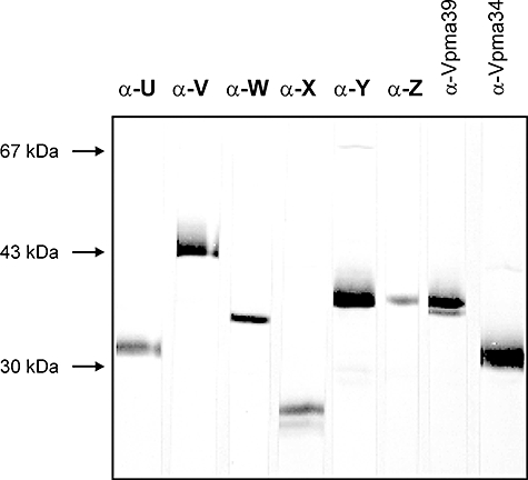
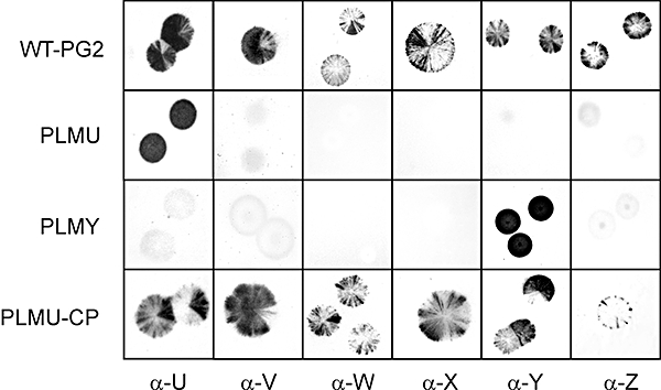
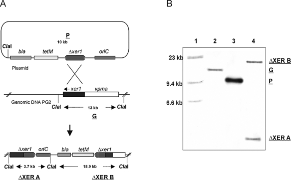
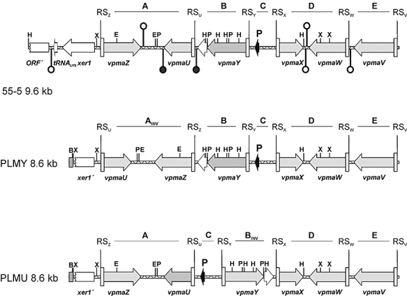
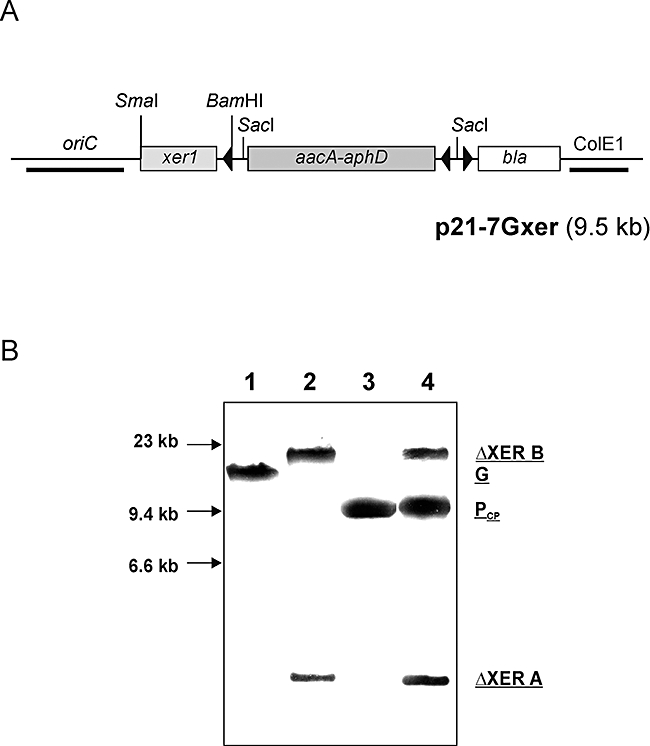
Mycoplasma agalactiae, an important pathogen of small ruminants, exhibits antigenic diversity by switching the expression of multiple surface lipoproteins called Vpmas (Variable proteins of M. agalactiae). Although phase variation has been shown to play important roles in many host-pathogen interactions, the biological significance and the mechanism of Vpma oscillations remain largely unclear. Here, we demonstrate that all six Vpma proteins are expressed in the type strain PG2 and all undergo phase variation at an unusually high frequency. Furthermore, targeted gene disruption of the xer1 gene encoding a putative site-specific recombinase adjacent to the vpma locus was accomplished via homologous recombination using a replicon-based vector. Inactivation of xer1 abolished further Vpma switching and the 'phase-locked' mutants (PLMs) continued to steadily express only a single Vpma product. Complementation of the wild-type xer1 gene in PLMs restored Vpma phase variation thereby proving that Xer1 is essential for vpma inversions. The study is not only instrumental in enhancing our ability to understand the role of Vpmas in M. agalactiae infections but also provides useful molecular approaches to study potential disease factors in other 'difficult-to-manipulate' mycoplasmas.


Figures

Fig. 1
Fig. 1
Western blot analyses of Triton X-114 phase material from M. agalactiae type strain PG2 using pAbs α-U to α-Z (as described in Table 1). Rabbit pAbs α-Vpma39 and α-Vpma34, previously raised against denatured VpmaY and VpmaU epitopes (Glew et al., 2000), respectively, served as positive controls. Protein size standards are indicated in the left margin.
Fig. 2
Fig. 2
Colony immunoblot analysis of M. agalactiae type strain PG2 and its derivative mutants using the six Vpma-specific pAbs recognizing specific surface exposed epitopes. PLMU and PLMY represent the two xer1-disrupted PLMs expressing exclusively VpmaU and VpmaY respectively. WT-PG2 and PLMU-CP representing the xer1-complemented PLMU show sectorial staining phenotype with all Vpma-specific pAbs reflecting high frequency Vpma phase variations. Designations of Vpma-specific pAbs as used for each column, and as described in Table 1, are indicated at the bottom.
Fig. 3
Fig. 3
Disruption of xer1 recombinase in M. agalactiae. A. Schematic representation of the integration of disruption plasmid pR3 (P) into the genomic DNA of type strain PG2 to generate the PLMU and PLMY clones. A 13 kb ClaI fragment of PG2 (G) consists of the known 9.6 kb vpma locus including the six vpma genes (white region) and the complete xer1 gene (black shaded region). A single putative homologous recombination event between the partial xer1 sequence carried by the plasmid pR3 and the chromosomal xer1 region is represented by crossed lines. This crossing over would lead to the integration of pR3 into the chromosome and would segregate the xer1 region onto two ClaI fragments ΔXERA and ΔXERB. B. Southern blot hybridization showing the localization of pR3 at the chromosomal xer1 locus of M. agalactiae PG2. ClaI-digested DNA of xer1 disruptant (PLMU or PLMY) (lane 4), disruption plasmid pR3 (lane 3) and wt strain PG2 (lane 2) were probed with a xer1-specific DIG-labelled fragment. λ-HindIII DNA size marker (lane 1).
Fig. 4
Fig. 4
Comparative Western blot analysis of whole-cell extracts of M. agalactiae type strain PG2 (WT-PG2), two PLMs (PLMY and PLMU) and xer1-complemented PLMU (PLMU-CP) using six different Vpma-specific pAbs as described in Table 1. Designations of individual pAbs used for each Western blot are indicated in the right margins of each panel whereas relevant protein size standards are shown on the left margins.
Fig. 5
Fig. 5
Arrangement of the ORFs, terminator structures and inversions in the vpma loci of M. agalactiae type strain PG2 clonal variant 55-5, PLMY and PLMU. Grey arrows represent the vpma genes, whereby a dark grey arrow denotes the vpma gene which is transcribed in the respective clonal variant or PLM. The black arrows indicate the location of the unique promoter (P) in each vpma locus. Putative recombination sites are indicated by white rectangles (RS) and denote sequences from −1 to −72 relative to the start codons of the corresponding vpma genes. White arrows indicate ORFs other than vpma genes, like the intact xer1 recombinase (xer1) and a tRNA (tRNALYS) gene in 55-5, whereas the discontinued white rectangles represent the promoter proximal regions of the disrupted xer1 genes (xer′) in the PLMs and a partial ORF (ORF′) in 55-5. A small grey rectangle indicates sequences derived from the integrated plasmid pR3. Non-translated repeats of the vpmaY genes are indicated with a dotted-lined white arrow. Terminator structures are indicated with symbolized hairpins, whereby Rho-independent terminator structures, for which experimental evidence is given, are drawn with a black-filled loop and are white-filled otherwise. Sequence-identical blocks between PLMs and 55-5 are designated A to E and are indicated with INV when inverted compared with 55-5. B, BamHI; E, EcoRI; H, HindIII; P, PstI; X, XbaI.
Fig. 6
Fig. 6
Complementation of the wt xer1 gene in PLMU. A. Schematic representation of complementation plasmid p21-7Gxer. Restriction sites used for cloning purposes are as indicated and arrowheads represent direction of transcription; ColE1, E. coli origin of replication; bla, ampicillin resistance gene; aacA-aphD, GentR gene; oriC, M. agalactiae origin of replication. B. Southern blot analysis of xer1-complemented PLMU. ClaI digest of DNA from the complementation clone PLMU-CP (lane 4) is shown in comparison with wt strain PG2 (lane 1), PLMU (lane 2) and complementation plasmid p21-7Gxer (lane 3) after hybridization with a xer1-specific DIG-labelled probe. G represents the 13 kb ClaI fragment of M. agalactiae type strain PG2 whereas ΔXERA and ΔXERB represent the two ClaI fragments from PLMU (as described in Fig. 3); PCP depicts the 9.5 kb linearized complementation plasmid p21-7Gxer. DNA size standards are indicated in the left margin.

Similar articles

Cited by

References

    1. Adams CD, Schnurr B, Skoko D, Marko JF, Reznikoff WS. Tn5 transposase loops DNA in the absence of Tn5 transposon end sequences. Mol Microbiol. 2006;62:1558–1568. - PubMed
    1. Altenbuchner J, Schmitt R. Transposon Tn1721: site-specific recombination generates deletions and inversions. Mol Gen Genet. 1983;190:300–308. - PubMed
    1. Altschul SF, Madden TL, Schaffer AA, Zhang J, Zhang Z, Miller W, Lipman DJ. Gapped blast and psi-blast: a new generation of protein database search programs. Nucleic Acids Res. 1997;25:3389–3402. - PMC - PubMed
    1. Askaa G, Erno H. ) Elevation of Mycoplasma agalactiae subsp. bovis to species rank: Mycoplasma bovis (Hale et al.) comb. nov. Int J Syst Bacteriol. 1976;26:323–325.
    1. d'Aubenton Carafa Y, Brody E, Thermes C. Prediction of rho-independent Escherichia coli transcription terminators. A statistical analysis of their RNA stem-loop structures. J Mol Biol. 1990;216:835–858. - PubMed

Publication types