Comparative Study
. 2008 Jun;105(5):1901-14.
doi: 10.1111/j.1471-4159.2008.05275.x. Epub 2008 Feb 4.

HIF-1 alpha is an essential effector for purine nucleoside-mediated neuroprotection against hypoxia in PC12 cells and primary cerebellar granule neurons

Affiliations
Comparative Study

HIF-1 alpha is an essential effector for purine nucleoside-mediated neuroprotection against hypoxia in PC12 cells and primary cerebellar granule neurons

Stephanie zur Nedden et al. J Neurochem. 2008 Jun.

Abstract

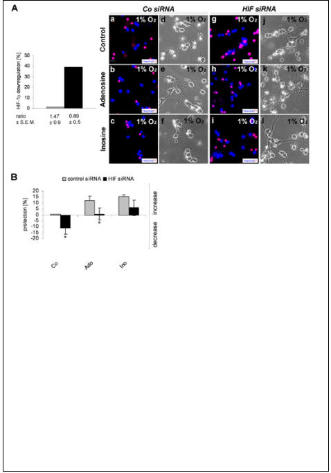
Hypoxia-inducible factor-1 alpha (HIF-1alpha) and purine nucleosides adenosine and inosine are critical mediators of physiological responses to acute and chronic hypoxia. The specific aim of this paper was to evaluate the potential role of HIF-1alpha in purine-mediated neuroprotection. We show that adenosine and inosine efficiently rescued clonal rat pheochromocytoma (PC12) cells (up to 43.6%) as well as primary cerebellar granule neurons (up to 25.1%) from hypoxic insult, and furthermore, that HIF-1alpha is critical for purine-mediated neuroprotection. Next, we studied hypoxia or purine nucleoside increased nuclear accumulation of HIF-1alpha in PC12 cells. As a possible result of increased protein stabilization or synthesis an up to 2.5-fold induction of HIF-1alpha accumulation was detected. In cerebellar granule neurons, purine nucleosides induced an up to 3.1-fold HIF-1alpha accumulation in cell lysates. Concomitant with these results, small interfering RNA-mediated reduction of HIF-1alpha completely abolished adenosine- and inosine-mediated protection in PC12 cells and severely hampered purine nucleoside-mediated protection in primary neurons (up to 94.2%). Data presented in this paper thus clearly demonstrate that HIF-1alpha is a key regulator of purine nucleoside-mediated rescue of hypoxic neuronal cells.


Figures

Figure 1
Figure 1
Effect of purine nucleosides on the cell viability and neurite outgrowth of hypoxic PC12 cells: Cells were exposed to normoxia and hypoxia (1% oxygen) for 24 hours in serum-reduced medium. Prior to hypoxic insult the cells were treated without purine nucleosides (Co) or with 500μM adenosine (Ado) and inosine (Ino). (A) Cells were stained with Hoechst (blue) and propidium iodide (PI, pink). (B) The percent of cell death is shown for normoxia (grey) and hypoxia (black). (C) The percent of cells protected from hypoxia by purine nucleosides is compared to the percent of cell death in controls (Co). (D) Representative phase contrast pictures of PC12 cells are shown for each experimental condition. (E) The percent of neurite outgrowth is shown for normoxia (grey) and hypoxia (black). (F) The fold increase in neurite outgrowth over control during hypoxia is shown for adenosine and inosine. Values represent the mean ± S.E.M., n=8-11. Differences were analyzed using unpaired one-tailed t-test: (B) ***p<0.001 (C) **p<0.01, ***p<0.001, (E) ***p<0.001, (F) **p<0.01. Scale bars: (A, D) 20μm.
Figure 2
Figure 2
Effect of purine nucleosides on cell viability of hypoxic cerebellar granule neurons: Cerebellar granule neurons from 7 day old rats were isolated and after 3 days in culture cells were exposed to normoxia and hypoxia (1% oxygen) for 6-8 hours. Prior to hypoxic insult cells were treated without purine nucleosides (Co) or with 500μM adenosine (Ado) and inosine (Ino). (A) Cells were stained with Hoechst (blue) and propidium iodide (PI, pink). (B) The percent of cell death is shown for normoxia (grey) and hypoxia (black). (C) The percent of cells protected from hypoxia by purine nucleosides is compared to the percent of cell death in controls (Co). Values represent the mean ± S.E.M., n=4. Differences were analyzed using unpaired one-tailed t-test: (B) *p<0.05 , (C) ***p<0.001. Scale bar: (A) 20 μm.
Figure 3
Figure 3
Effect of purine nucleosides on HIF-1α protein accumulation, and DNA-binding capacity in PC12 cells: PC12 cells were exposed to normoxia and hypoxia (1% oxygen) for 24 hours in serum-reduced medium. Prior to hypoxic insult cells were treated without purine nucleosides (Co) or with 500μM adenosine (Ado) and inosine (Ino). HIF-1α protein levels of (A) total cell extracts and (B) nuclear extracts were determined by western blot analysis. DNA polymerase was used as a loading control and the ratios of HIF-1α/DNA polymerase are shown for normoxia (grey) and hypoxia (black) in control cells (Co) in panels Aa and Ba. Nuclear extracts were also treated with CoCl2 (positive control). Levels of hypoxic HIF-1α were the basis for calculation of the additional purine-mediated increase (Ab and Bb). (C) Nuclear accumulation of HIF-1α was tested by immunofluorescence analysis. (Panels a-e) Hoechst stain (blue) was used to visualize the nucleus of the cells; (Panels f-j). An FITC-labeled secondary antibody (green) was used to visualize HIF-1α, and (panels k-o) the same cells are shown under phase contrast optics. (C Panels a, f and k) CoCl2-treated samples are shown for comparison. (D) The DNA-binding capacity of nuclear extracts was analyzed by EMSA kit. The colorimetric readout was quantified by spectrophotometry. Fold increases over control (Co) indicate increases in nuclear HIF-1α binding to immobilized DNA. Values represent the mean ± S.E.M., n=3-9. Differences were analyzed using unpaired one-tailed t-test: (A) **p<0.01, ***p<0.001, (B) *p<0.05. Scale bar: (C) 20μm.
Figure 4
Figure 4
Effect of purine nucleosides on HIF-1α protein accumulation in cerebellar granule neurons: Cerebellar granule neurons from 7 day old rats were isolated and after 3 days, cells were exposed to hypoxia (1% oxygen) for 6-8 hours. Prior to hypoxic insult cells were treated without purine nucleosides (Co) or with 500μM adenosine (Ado) and inosine (Ino). (A) The HIF-1α protein level in total cell extracts was determined by western blot analysis. DNA polymerase was used as a loading control, and the ratios of HIF-1α/DNA polymerase are shown for normoxia (grey) and hypoxia (black) in control cells (Co) in panel Aa and for purine-treated cells in panel Ab. (B) Nuclear accumulation of HIF-1α was tested by immunofluorescence analysis. (Panels a-d) Hoechst stain (blue) was used to visualize the nucleus of the cells; (Panels e-h) An FITC-labeled secondary antibody (green) was used to visualize HIF-1α. Values represent the mean ± S.E.M., n=5. Differences were analyzed using unpaired one-tailed t-test: (A) *p<0.05. Scale bar: (B) 20μm.
Figure 5
Figure 5
Effect of partial HIF-1α knockdown in PC12 cells: PC12 cells were transfected with siRNA directed against HIF-1α and a non-targeting siRNA pool, which was used as control (control siRNA). After 2 days in culture cells were exposed to hypoxia (1% oxygen) for 24 hours in serum-reduced medium. Prior to hypoxic insult cells were treated without purine nucleosides (Co) or with 500μM adenosine (Ado) and inosine (Ino). (A) Panels a-f indicate cell death under hypoxia in cells transfected with control and HIF-1α siRNA. Cells were stained with Hoechst (blue) and propidium iodide (PI, pink). (Left panel) The percent of HIF-1α downregulation is shown for cells transfected with control siRNA (grey) and HIF-1α siRNA (black). (g) The percent of cells protected from hypoxia by purine nucleosides is shown for cells transfected with HIF-1α siRNA and control siRNA. (B) Representative phase contrast pictures show neurite outgrowth in cells transfected with HIF-1α and control siRNA. Panel g shows the fold changes in neurite outgrowth under the various conditions. Values were normalized to the neurite outgrowth levels observed in hypoxic cells transfected with control siRNA without nucleosides. Values represent the mean ± S.E.M., n=4. Differences were analyzed using unpaired one-tailed t-test: (A) *p<0.05, **p=0.01, (B) **p<0.01. Scale bars: (A, B) 20μm.
Figure 6
Figure 6
Effect of partial HIF-1α knockdown in cerebellar granule neurons: Cerebellar granule neurons were transfected with siRNA directed against HIF-1α and a non-targeting siRNA pool, which was used as control (control siRNA). After 3 days in culture cells were exposed to hypoxia (1% oxygen) for 6-8 hours. Prior to hypoxic insult cells were treated without purine nucleosides (Co) or with 500μM adenosine (Ado) and inosine (Ino) respectively. (A) Panels a-c and g-i indicate cell death in hypoxia in cells transfected with control and HIF-1α siRNA. Cells were stained with Hoechst (blue) and propidium iodide (PI, pink). Panels d-f and j-l show representative phase contrast pictures of the same cells. (Left panel) The percent of HIF-1α downregulation is shown for cells transfected with control siRNA (grey) and HIF-1α siRNA (black). (B) The percent of cells protected from hypoxia by purine nucleosides is shown for cells transfected with HIF-1α and control siRNA. Values represent the mean ± S.E.M., n=6. Differences were analyzed using unpaired one-tailed t-test: (B) *p<0.05. Scale bar: (A) 20μm.

Similar articles

Cited by

References

    1. Arslan G, Kull B, Fredholm BB. Signaling via A2A adenosine receptor in four PC12 cell clones. Naunyn Schmiedebergs Arch Pharmacol. 1999;359:28–32. - PubMed
    1. Ballarin M, Fredholm BB, Ambrosio S, Mahy N. Extracellular levels of adenosine and its metabolites in the striatum of awake rats: inhibition of uptake and metabolism. Acta Physiol Scand. 1991;142:97–103. - PubMed
    1. Baranova O, Miranda LF, Pichiule P, Dragatsis I, Johnson RS, Chavez JC. Neuron-specific inactivation of the hypoxia inducible factor 1alpha increases brain injury in a mouse model of transient focal cerebral ischemia. J Neurosci. 2007;27:6320–6332. - PMC - PubMed
    1. Batistatou A, Volonté C, Greene LA. Nerve growth factor employs multiple pathways to induce primary response genes in PC12 cells. Mol Biol Cell. 1992;3:363–371. - PMC - PubMed
    1. Benowitz LI, Jing Y, Tabibiazar R, Jo SA, Petrausch B, Stuermer CA, Rosenberg PA, Irwin N. Axon outgrowth is regulated by an intracellular purine-sensitive mechanism in retinal ganglion cells. J Biol Chem. 1998;273:29626–29634. - PubMed

Publication types

Substances