Comparative Study
. 2008 Jun;105(5):1770-80.
doi: 10.1111/j.1471-4159.2008.05272.x. Epub 2008 Feb 4.

N,N-dimethyl-thioamphetamine and methyl-thioamphetamine, two non-neurotoxic substrates of 5-HT transporters, have scant in vitro efficacy for the induction of transporter-mediated 5-HT release and currents

Affiliations
Comparative Study

N,N-dimethyl-thioamphetamine and methyl-thioamphetamine, two non-neurotoxic substrates of 5-HT transporters, have scant in vitro efficacy for the induction of transporter-mediated 5-HT release and currents

Marco Gobbi et al. J Neurochem. 2008 Jun.

Abstract

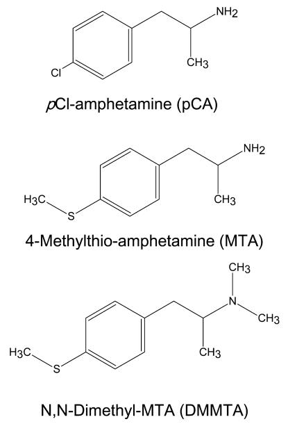
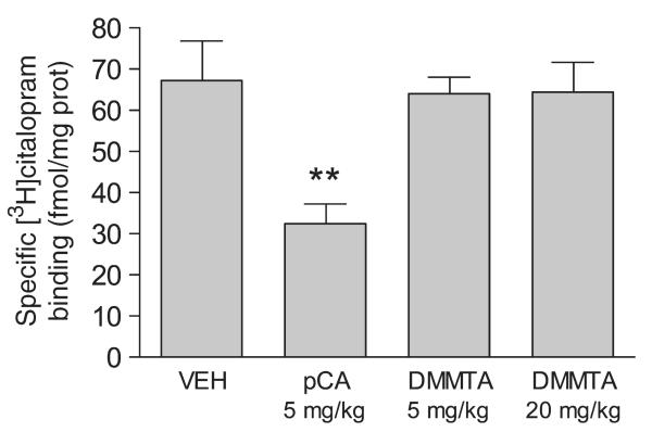
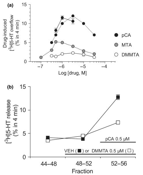
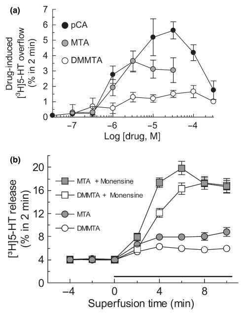
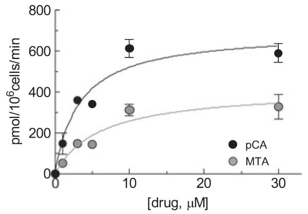
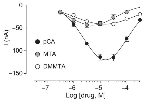
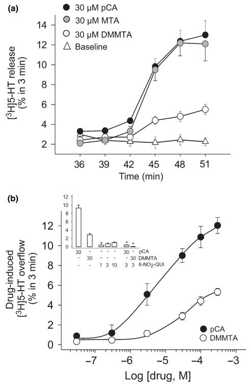
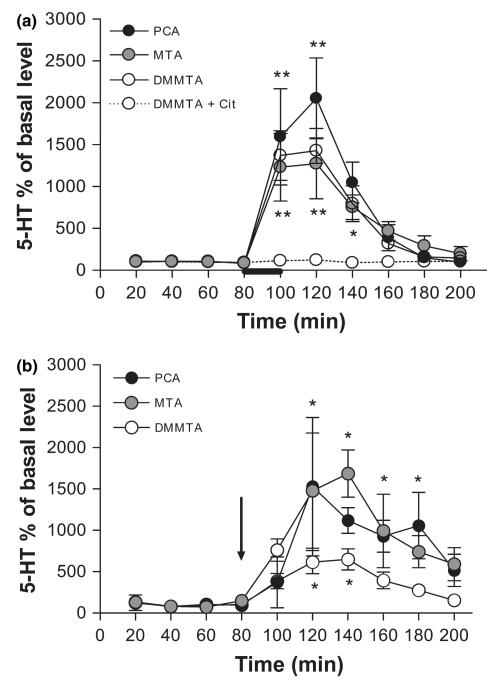
We studied two non-neurotoxic amphetamine derivatives (methyl-thioamphetamine, MTA and N,N-dimethylMTA, DMMTA) interacting with serotonin (5-HT) transporters (SERTs) with affinities comparable to that of p-Cl-amphetamine (pCA). The rank order for their maximal effects in inducing both [(3)H]5-HT release from rat brain synaptosomes or hSERT-expressing HEK-293 cells, and currents in hSERT-expressing oocytes, was pCA >> MTA > or = DMMTA. A correlation between drug-induced release and currents is also strengthened by the similar bell shape of the dose-response curves. Release experiments indicated that MTA and DMMTA are SERT substrates although MTA is taken up by HEK-293 cells with a V(max) 40% lower than pCA. The weak effects of MTA and DMMTA in vitro might therefore be due to their properties as 'partial substrates' on the mechanisms, other than translocation, responsible for currents and/or release. After either local or systemic in vivo administration, MTA and DMMTA release 5-HT in a manner comparable to pCA. These findings confirm that the neurotoxic properties of some amphetamine derivatives are independent of their 5-HT-releasing activity in vivo. It is worth noting that only those amphetamine derivatives with high efficiency in inducing 5-HT release and currents in vitro have neurotoxic properties.


Figures

Fig. 1
Fig. 1
Chemical structures of the amphetamine derivatives used in the present study.
Fig. 2
Fig. 2
In vivo treatment with DMMTA was not neurotoxic for 5-HTergic neurons in rat hippocampus. Male rats received one intraperitoneal injection of 5 or 20 mg/kg DMMTA or 5 mg/kg pCA. Control rats received vehicle alone (saline solution). One week later, rats were killed and [3H]citalopram binding was measured in their hippocampi. Each value represents the mean ± SE of five rats. **p < 0.01 versus vehicle-treated rats.
Fig. 3
Fig. 3
Panel (a). Releasing effect of DMMTA from superfused rat hippocampal synaptosomes preloaded with [3H]5-HT. Release was calculated by subtracting the fractional release rate (FRR) immediately before the stimulus (basal release) from that immediately after. Each value is the mean ± SEM of 3–12 replications from 1 to 4 experiments; in most cases SEM is within the size of the symbols. In each experiment testing DMMTA derivatives, pCA was always added as a positive internal control and the results are shown. Dashed lines show the concentration-effect curves previously reported with pCA and MTA (Gobbi et al. 2002), for comparison. Panel (b) DMMTA (0.5 μM) antagonized the [3H]5-HT release induced by 0.5 μM pCA. Synaptosomes were exposed to DMMTA, or its vehicle (VEH) from t = 48 min onward and pCA was added from t = 52 min. Three 4-min fractions were collected: t = 44–48 (basal release), t = 48–52 (DMMTA-induced release) and t = 52–56 (pCA-induced release in the absence or presence of DMMTA). Ordinates indicate the [3H]5-HT released in each 4-min fraction as a percentage of the total radioactivity in the synaptosomes at the start of that fraction. Each value is the mean ± SEM of four replications (four chambers in parallel). Two-way anova was done using the data of fractions 2 and 3, and the results indicated significant interactions (p < 0.01) between DMMTA and pCA.
Fig. 4
Fig. 4
Panel (a) Releasing effect of DMMTA, MTA, and pCA from superfused SERT-expressing HEK-293 cells preloaded with [3H]5-HT. Drug-induced release is the difference between the mean fractional release rate (FRR) after and before cells are exposed to drugs. Symbols represent mean ± SEM of 6–9 observations from two or three independent experiments. Panel (b) Effect of monensin on the [3H]5-HT-releasing effects of MTA and DMMTA. After three fractions of basal efflux, the cells were exposed, as indicated, to 10 μM MTA or DMMTA, in the absence or presence of 10 μM monensin. Data are presented as FRR. Symbols represent mean ± SEM of nine observations from three separate experiments.
Fig. 5
Fig. 5
Concentration dependence of uptake of MTA and pCA in HEK-293 cells stably expressing hSERT. Cells were incubated for 1 min with MTA and pCA at the concentrations indicated. Amphetamines were then extracted from the cells and determined by HPLC as described under Materials and methods. Symbols show the specific (i.e., paroxetine-sensitive) uptake and are means ± SEM of three experiments run in triplicate. The saturation curves were fitted using the ‘one-site model’ hyperbola equation (GraphPad Prism 4.0a). The calculated Vmax of MTA (416 ± 58) was significantly lower (p < 0.01) than the Vmax of pCA (695 ± 58) whereas Km (6.0 ± 2.3 vs. 3.3 ± 1.0 μM, respectively) were not significantly different (compared with F-tests included in the GraphPad Prism 4.0a software).
Fig. 6
Fig. 6
Inward currents induced by application of different concentrations of pCA, DMMTA and MTA to hSERT expressing Xenopus laevis oocytes. Electrophysiological experiments were done using the two-electrode voltage-clamp technique with oocytes clamped to a holding potential of −80 mV. Data are means ± SEM of three experiments performed in triplicate.
Fig. 7
Fig. 7
Releasing effect of DMMTA from rat brain hippocampal slices preloaded with [3H]5-HT. Slices were prepared, labeled with the radioactive tracer and exposed to drugs in superfusion. DMMTA, MTA or pCA were added at t = 39 min of superfusion and maintained until the end of the experiments. 6-Nitro-quipazine (6-NO2-QUI) was introduced at t = 30 min. Panel (a) Time course of the release of [3H]5-HT from slices exposed to 30 μM DMMTA, MTA or pCA. Data are expressed as FRR (see Materials and methods). Panel (b) and inset. Concentration–response curve of the effects of DMMTA or pCA and block by 6-NO2-QUI. Data are expressed as drug-induced [3H]5-HT overflow, calculated by subtracting the basal release (second fraction collected; t = 36–39 min) from the release measured in the fifth fraction collected (t = 45–48 min), where the drugs generally reached maximum effect. The data presented are mean ± SEM of 4–6 experiments in triplicate. *p < 0.001 compared to the effect of pCA or DMMTA alone (two-tailed Student’s t-test).
Fig. 8
Fig. 8
Microdialysis data showing the effects of DMMTA, MTA or pCA either applied locally (panel a, drugs infused at a concentration of 1 mM as indicated by the bar) or given systemically (panel b, i.p. injection of 5 mg/kg is indicated by the arrow) on extracellular 5-HT in the rat lateral septum (LS) and in the dorsal hippocampus. Data are mean ± SEM (n = 4–6 for each condition). Asterisks indicate a significant difference between samples after treatment and the corresponding basal values, calculated by one-way anova-Student–Newman–Keuls: *p < 0.05, and **p < 0.01.

References

    1. Baumann MH, Ayestas MA, Dersch CM, Rothman RB. 1-(m-chlorophenyl)piperazine (mCPP) dissociates in vivo serotonin release from long-term serotonin depletion in rat brain. Neuropsychopharmacology. 2001;24:492–501. - PubMed
    1. Crespi D, Mennini T, Gobbi M. Carrier-dependent and Ca(2 + )-dependent 5-HT and dopamine release induced by (+)-amphetamine, 3,4-methylendioxymethamphetamine, p-chloroamphetamine and (+)-fenfluramine. Br. J. Pharmacol. 1997;121:1735–1743. - PMC - PubMed
    1. Dinopoulos A, Dori I, Parnavelas JG. Serotonergic innervation of the mature and developing lateral septum of the rat: a light and electron microscopic immunocytochemical analysis. Neuroscience. 1993;55:209–222. - PubMed
    1. Eriksson E, Engberg G, Bing O, Nissbrandt H. Effects of mCPP on the extracellular concentrations of serotonin and dopa-mine in rat brain. Neuropsychopharmacology. 1999;20:287–296. - PubMed
    1. Fischer JF, Cho AK. Chemical release of dopamine from striatal homogenates: evidence for an exchange diffusion model. J. Pharmacol. Exp. Ther. 1979;208:203–209. - PubMed

Publication types

MeSH terms