. 2008 Jun;10(6):1286-303.
doi: 10.1111/j.1462-5822.2008.01126.x. Epub 2008 Jan 31.

The Delta fbpA mutant derived from Mycobacterium tuberculosis H37Rv has an enhanced susceptibility to intracellular antimicrobial oxidative mechanisms, undergoes limited phagosome maturation and activates macrophages and dendritic cells

Affiliations

The Delta fbpA mutant derived from Mycobacterium tuberculosis H37Rv has an enhanced susceptibility to intracellular antimicrobial oxidative mechanisms, undergoes limited phagosome maturation and activates macrophages and dendritic cells

Muralidhar K Katti et al. Cell Microbiol. 2008 Jun.

Abstract

Mycobacterium tuberculosis H37Rv (Mtb) excludes phagocyte oxidase (phox) and inducible nitric oxide synthase (iNOS) while preventing lysosomal fusion in macrophages (MPhis). The antigen 85A deficient (Delta fbpA) mutant of Mtb was vaccinogenic in mice and the mechanisms of attenuation were compared with MPhis infected with H37Rv and BCG. Delta fbpA contained reduced amounts of trehalose 6, 6, dimycolate and induced minimal levels of SOCS-1 in MPhis. Blockade of oxidants enhanced the growth of Delta fbpA in MPhis that correlated with increased colocalization with phox and iNOS. Green fluorescent protein-expressing strains within MPhis or purified phagosomes were analysed for endosomal traffick with immunofluorescence and Western blot. Delta fbpA phagosomes were enriched for rab5, rab11, LAMP-1 and Hck suggesting enhanced fusion with early, recycling and late endosomes in MPhis compared with BCG or H37Rv. Delta fbpA phagosomes were thus more mature than H37Rv or BCG although, they failed to acquire rab7 and CD63 preventing lysosomal fusion. Finally, Delta fbpA infected MPhis and dendritic cells (DCs) showed an enhanced MHC-II and CD1d expression and primed immune T cells to release more IFN-gamma compared with those infected with BCG and H37Rv. Delta fbpA was thus more immunogenic in MPhis and DCs because of an enhanced susceptibility to oxidants and increased maturation.


Figures

Fig. 1
Fig. 1
Disruption of fbpA gene leads to reduced cell wall lipids in ΔfbpA mutant compared with wild-type Mycobacterium tuberculosis (Mtb, H37Rv) and reconstituted ΔfbpA. Bacteria were grown in the presence of 14C-acetate in 7H9 broth and at hourly intervals thin layer chromatographic analysis of cell wall extracts was performed followed by radiography. The bars represent c.p.m. ± SD (not apparent) of two similar experiments. Wild-type H37Rv incorporates more label than ΔfbpA mutant and the reconstitution of the gene restores the incorporation of radiolabel into the mutant (P-values shown for strains compared; Student's t-test).
Fig. 2
Fig. 2
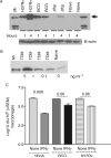
ΔfbpA affects signal transduction in MΦs through the delayed induction of suppressor of cytokine signalling 1 (SOCS-1). A. ΔfbpA induces less strong SOCS-1 response. C57BL/6 mouse bone marrow-derived macrophages (MΦs) were infected with mycobacteria at an moi of 1:1 and incubated for 1 and 4 h. MΦ lysates were analysed using 7–12% SDS gels and electroblotted membranes were probed with an antibody to SOCS-1, followed by HRP-conjugate. Bands were detected using ECL. H37Rv and BCG induce copious expression of SOCS-1 in MΦs 1 and 4 h after infection. ΔfbpA does not induce SOCS-1 at 1 h but relatively lower levels of SOCS-1 are apparent at 4 h post infection (Mr, molecular weight marker). Lanes were loaded with equal amounts of protein as indicated by β-actin control. B. Purified trehalose 6,6, dimycolate (TDM) induces SOCS-1. Varying amounts of TDM suspension in mineral oil were added to MΦs and lysates examined at 1 h after treatment for SOCS-1 as above. Naïve MΦs were treated with mineral oil control. C. ΔfbpA is more susceptible to IFN-γ. MΦs were infected with mycobacteria (moi 1:1), washed to remove extracellular bacteria and 4 h later incubated with 400 U ml–1 of IFN-γ. 48 h later, the MΦs were lysed and plated for colony-forming unit (cfu) counts on 7H11 agar. Data summarizing three separate experiments show that IFN-γ is more effective in reducing the cfu of ΔfbpA when compared with its effects on MΦs infected with either BCG or H37Rv (P-values shown for groups compared; Student's t-test).
Fig. 3
Fig. 3
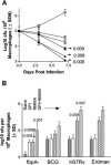
Growth and susceptibility of ΔfbpA mutant, H37Rv and BCG vaccine to intracellular oxidants. A. Untreated (open symbols) or IFN-γ activated MΦs (closed symbols; 400 U ml–1 for 24 h) were infected with the three strains of mycobacteria and incubated. On days shown, triplicate wells of MΦs per strain were lysed and plated for cfu on 7H11 agar in three separate experiments. ΔfbpA (Δ) and BCG (○) are unable to grow while the wild-type H37Rv (□) shows an increase in cfu counts over 7 days. IFN-γ activation of MΦs decreases the cfu of all three strains by more than 1.5 log10 (P-values for growth in naïve MΦs versus IFN-γ activated MΦs; Student's t-test). B. MΦs were infected as above and cfu determined on day 3 (see Results) in the presence or absence of an inhibitor of reactive oxygen species (ROS; diphenylene iodonium, DPI) or nitric oxide (NO) synthesis (NG-monomethyl-l-arginine, NMMA) or their combination. DPI and NMMA enhance the growth of ΔfbpA. DPI is ineffective against H37Rv while NMMA is effective. BCG shows a modest increase in growth with a combination of DPI and NMMA (P-values are shown above the bars versus growth of mycobacteria in naïve MΦs; Student's t-test). MΦs infected with Erdman strain show a growth profile similar to H37Rv and was included as another positive control.
Fig. 4
Fig. 4
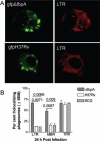
Phagosomes of ΔfbpA more frequently acquire inducible nitric oxide synthase (iNOS) and p47phox component of phagocyte oxidase in MΦs. MΦs were infected with green fluorescent protein (gfp) expressing mycobacterial strains (gfpDfbpA, gfpBCG and gfpH 37Rv) for 4 h, washed and incubated for 24 or 72 h. MΦs were then fixed, permeabilized and stained with a mouse monoclonal antibody to iNOS or goat anti-p47phox followed by anti-Ig-Texas red conjugates. MΦs were examined for colocalization using a Deltavision laser scanning microscope (LSM). Arrows illustrate colocalization in A and C. A. LSM profiles illustrate that gfpΔfbpA phagosomes acquire iNOS while gfpH 37Rv and gfpBCG acquire less. B. Per cent phagosomes positive for iNOS colocalization was tabulated from three experiments and P-values are shown for the difference between gfpDfbpA, gfpDBCG and gfpH 37Rv phagosomes (Student's t-test). At least 200 microscopic fields were scored blind per mouse MΦ preparation. Colocalizing bacteria were averaged from three independent experiments each with MΦs from three mice; each microscopic field contained about five MΦs, each of which contained about five bacilli. C. gfpΔfbpA phagosomes acquire more p47phox than either gfpH 37Rv or gfpBCG. D. Phagosomes positive for colocalization are shown; data averaged from three separate experiments show increased phox colocalization for gfpΔfbpA versus gfpDBCG and gfpH 37Rv.
Fig. 5
Fig. 5
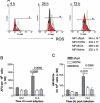
A. Intracellular oxidative activity within MΦs infected with ΔfbpA, BCG and H37Rv. MΦs or C57BL/6 bone marrow (BM)-derived BMA.A3 macrophage cell lines (both similar, MΦs shown) were infected with mycobacteria at an moi of 1:1, washed and incubated for 72 h. A. At time points shown, cells were treated with 1 μg ml–1 dihydrodichloro-fluorescein diacetate (H2DCFDA), a probe for intracellular reactive oxygen species (ROS) for 5 min, washed and analysed in BD-Facscan using Cellquest software. Viable naïve MΦs have a basal level of fluorescence indicated by a red fill. MΦs infected with ΔfbpA show an enhanced and sustained fluorescence over time (green). MΦs infected with H37Rv show minimal ROS activity (black) while BCG infected MΦs show an intermediate level (blue). A positive control of phorbol myristyl acetate (an agonist for ROS) activated MΦs showed a burst of activity that declines rapidly within minutes (not shown). Mean fluorescent intensity (MFI, inset) values for ΔfbpA calculated from triplicate experiments (one histogram illustrated) was significant using Student's t-test (*P < 0.0092). B and C. Fluorometry and colorimetry for oxidant activity in MΦs. MΦs were infected as above. Twenty-four and 72 h post infection, the cultures from triplicate experiments were tested for ROS using H2DCFDA and fluorometry (B) and nitrite, a product of NO synthesis by MΦs using the Greiss reagent and colorimetry (C). ΔfbpA induces accumulation of higher levels of ROS and nitrite and P-values are shown for gfpΔfbpA versus gfpΔBCG and gfpH 37Rv (Student's t-test). Background fluorescence for ROS or nitrite in uninfected MΦs is shown as a dotted line in both B and C.
Fig. 6
Fig. 6
ΔfbpA phagosomes efficiently acquire both early and late endosomal markers when compared with H37Rv and BCG within MΦs. MΦs were labelled with lysotracker red (LTR), mannosylated BSA rhodamine (MBR), or transferrin Texas red (TRR), chased with medium and infected with gfpΔfbpA, gfpBCG or gfpH 37Rv strains. MΦs were washed and examined for fluorescence colocalization. A. LSM profile illustrates that live gfpΔfbpA has markedly enhanced colocalization with LTR while the wild-type gfpH 37Rv excludes LTR. B. Per cent phagosomes positive for colocalization was determined by using LSM in three separate experiments. gfpΔfbpA significantly colocalizes with both LTR and MBR compared with either gfpBCG or gfpH 37Rv (Student's t-test). Colocalization with TRR, an early endosomal fusion marker, was comparable for all three strains tested. Scoring was carried out as in Fig. 4.
Fig. 7
Fig. 7
ΔfbpA phagosomes acquire late endosomal maturation markers but exclude lysosomal markers. MΦs were infected with gfpΔfbpA, gfpH37Rv and gfpBCG strains for 4 h, washed and incubated for 24 h. MΦs were fixed, permeabilized and stained for three markers of late endosomes (lysosome-associated membrane protein 1, LAMP-1; src family kinase, Hck) and lysosomes (CD63 and rab7) using specific antibodies and counterstained with anti-Ig-Texas red conjugates. Phagosomes were examined for colocalization (arrow and arrowhead). A. LSM profile illustrates that gfpΔfbpA has an enhanced colocalization (arrow) with the Hck. B. Illustration that gfpΔfbpA largely excludes rab7. C. Per cent phagosomes positive for fluorescence colocalization with the markers was determined by using LSM. gfpΔfbpA phagosomes are more enriched for LAMP-1 and Hck but not CD63 and rab7. gfpBCG and gfpH 37Rv also acquire LAMP-1 to a lower level but exclude Hck, CD63 and rab7. Scoring from three separate experiments was averaged and significance determined using Student's t-test. D. MΦs were infected with mycobacteria at an moi of 1:1 and phagosomes were purified on sucrose gradients at 24 h post infection. Latex beads (LB) were internalized for 4 h and phagosomes prepared as a positive control. Phagosomal proteins were then analysed using Western blot with specific antibodies followed by anti-Ig conjugates and HRP-dependent chemiluminescence. LB phagosomes acquire nearly all docking proteins along with CD63 and rab7, the specific marker of lysosomes. gfpΔfbpA phagosomes acquire multiple markers that suggest an ability to fuse with early and recycling endosomes such as rab5A, rab5B and rab11. While they acquire late endosomal markers such as Hck, they exclude lysosome-specific markers like CD63 and rab7. LAMP-1 and phosphatidyl inositol-3-kinase are also enriched on LB and gfpΔfbpA phagosomes compared with gfpBCG and gfpH 37Rv.
Fig. 8
Fig. 8
The ΔfbpA mutant induces an enhanced expression of MHC-II and CD1d molecules in MΦs and dendritic cells (DCs). A. MΦs or BM-derived BMA.A3 cell line MΦs (both showed similar results, MΦs shown) were infected with mycobacteria at an moi of 1:1 for 4 h, washed, incubated for 24 h and stained for surface receptor expression using specific antibodies conjugated to fluorochromes. Receptor expression was analysed using BD-Facscan and Cellquest software. MΦ histograms (one of three experiments illustrated) show the unstained or isotype stained cells (red fill); ΔfbpA (green); H37Rv (blue) and BCG (yellow) infected MΦs. Histograms illustrate that MΦs infected with ΔfbpA show an enhanced expression of CD40, CD86 and MHC-II when compared with BCG- or H37Rv-infected MΦs. Mean fluorescence intensity values (MFIs) from triplicate separate experiments are shown below the histograms (*P < 0.01; **P < 0.007; ***P < 0.008; #0.01 compared with H37Rv- or BCG-infected MΦs, Student's t-test). B. BM-derived CD11c bead purified DCs were infected and analysed for receptor expression as above. Histograms illustrate that ΔfbpA (blue) induces a stronger expression of MHC-II in DCs than BCG (black) or H37Rv (blue). MFIs from three independent experiments are shown below the histograms (*P < 0.009; **P < 0.009 compared BCG- or H37Rv-infected DCs, Student's t-test).
Fig. 9
Fig. 9
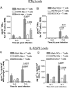
ΔfbpA is more immunogenic in MΦs or DCs than H37Rv or BCG. Antigen-presenting cells (APCS) such as MΦs (A, C) or DCs (B, D) were infected with mycobacteria for 4 h, washed and co-cultured with naïve or sensitized CD3-bead purified T cells from spleens of mice immunized with Mtb. After 72 h, the culture supernatants were assayed for IFN-γ (A and B) or IL-12p70 (C,D) using sandwich ELISA. A and B. ΔfbpA induces a stronger response of IFN-γ in immune T cells (shown as bars) than BCG or H37Rv within MΦs (A) or DCs (B) (P-values determined using Student's t-test). Neither naïve T cells co-cultured with infected APCs nor uninfected APCs secrete significant levels of IFN-γ. The IFN-γ levels for these controls are represented as horizontal dotted lines. C and D. ΔfbpA infected MΦs (C) or DCs (D) show elevated levels of IL-12p70 compared with BCG or H37Rv. Basal levels of IL-12p70 in uninfected in APCs or naïve T cell co-cultures are shown as horizontal dotted lines.

Similar articles

Cited by

References

    1. Armitige LY, Jagannath C, Wanger AR, Norris SJ. Disruption of the genes encoding antigen 85A and antigen 85B of Mycobacterium tuberculosis H37Rv: effect on growth in culture and in macrophages. Infect Immun. 2000;68:767–778. - PMC - PubMed
    1. Armstrong JA, Hart PD. Response of cultured macrophages to Mycobacterium tuberculosis, with observations on fusion of lysosomes with phagosomes. J Exp Med. 1971;134:713–740. - PMC - PubMed
    1. Astarie-Dequeker C, N'Diaye EN, Le Cabec V, Rittig MG, Prandi J, Maridonneau-Parini I. The mannose receptor mediates uptake of pathogenic and nonpathogenic mycobacteria and bypasses bactericidal responses in human macrophages. Infect Immun. 1999;67:469–477. - PMC - PubMed
    1. Astarie-Dequeker C, Carreno S, Cougoule C, Maridonneau-Parini I. The protein tyrosine kinase Hck is located on lysosomal vesicles that are physically and functionally distinct from CD63-positive lysosomes in human macrophages. J Cell Sci. 2002;115:81–89. - PubMed
    1. Babior BM. NADPH oxidase: an update. Blood. 1999;93:1464–1476. - PubMed

Publication types

MeSH terms

Substances

LinkOut - more resources