. 2008 Jun;7(3):335-43.
doi: 10.1111/j.1474-9726.2008.00377.x. Epub 2008 Jan 31.

Age-related intrinsic changes in human bone-marrow-derived mesenchymal stem cells and their differentiation to osteoblasts

Affiliations

Age-related intrinsic changes in human bone-marrow-derived mesenchymal stem cells and their differentiation to osteoblasts

Shuanhu Zhou et al. Aging Cell. 2008 Jun.

Abstract

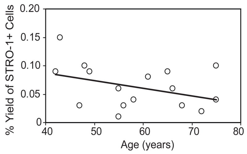
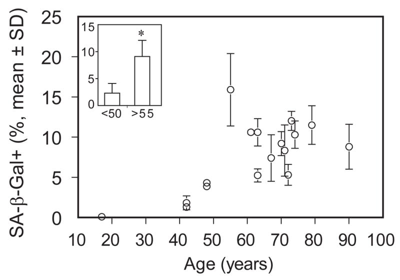
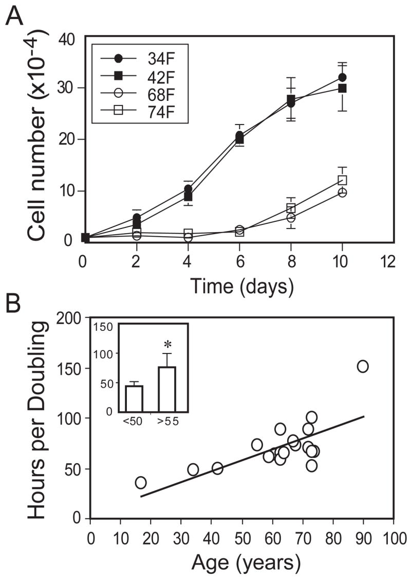
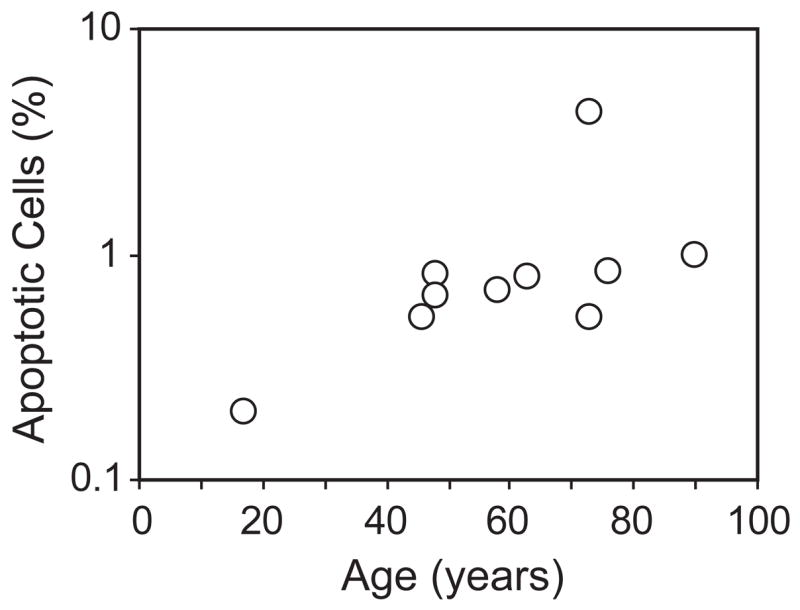
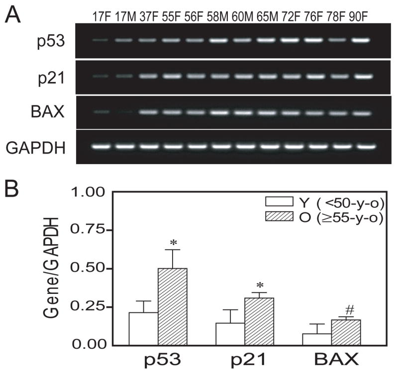
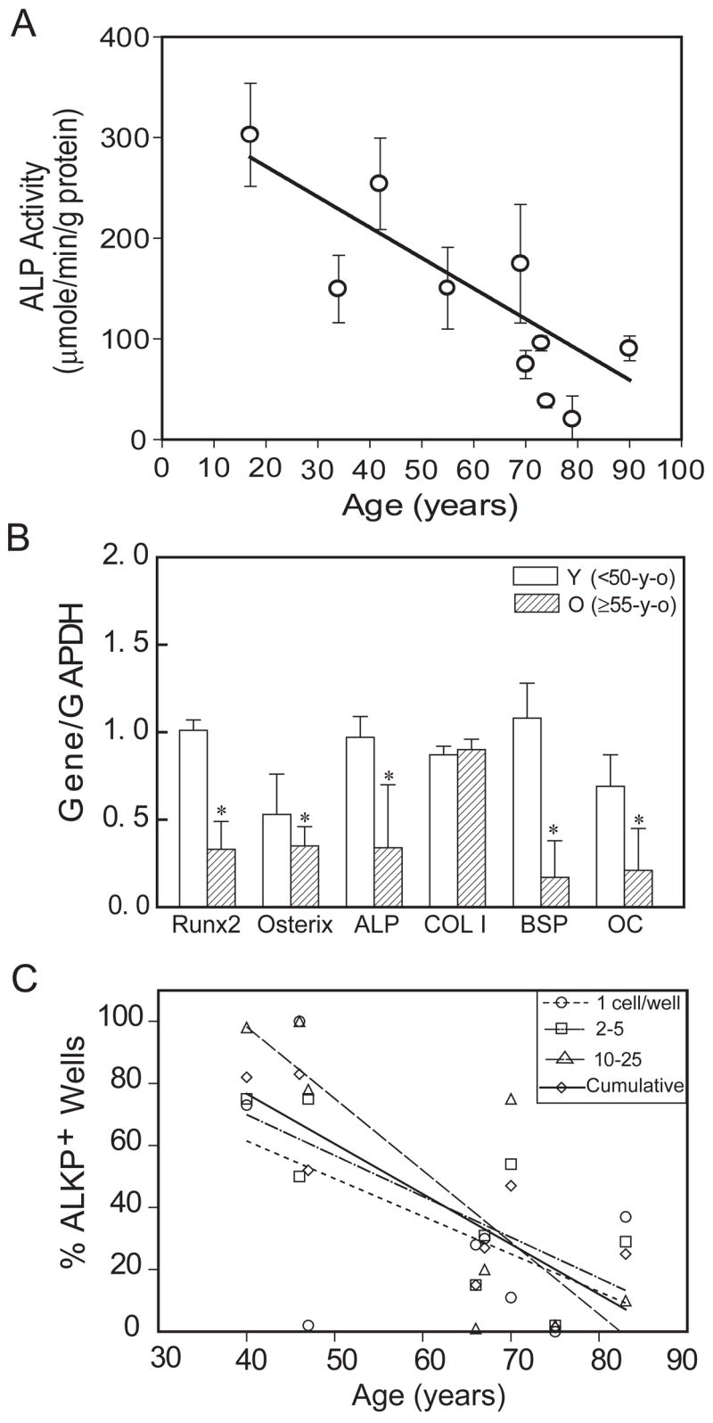
In vivo and in vitro studies indicate that a subpopulation of human marrow-derived stromal cells (MSCs, also known as mesenchymal stem cells) has potential to differentiate into multiple cell types, including osteoblasts. In this study, we tested the hypothesis that there are intrinsic effects of age in human MSCs (17-90 years). We tested the effect of age on senescence-associated beta-galactosidase, proliferation, apoptosis, p53 pathway genes, and osteoblast differentiation in confluent monolayers by alkaline phosphatase activity and osteoblast gene expression analysis. There were fourfold more human bone MSCs (hMSCs) positive for senescence-associated beta-galactosidase in samples from older than younger subjects (P < 0.001; n = 17). Doubling time of hMSCs was 1.7-fold longer in cells from the older than the younger subjects, and was positively correlated with age (P = 0.002; n = 19). Novel age-related changes were identified. With age, more cells were apoptotic (P = 0.016; n = 10). Further, there were age-related increases in expression of p53 and its pathway genes, p21 and BAX. Consistent with other experiments, there was a significant age-related decrease in generation of osteoblasts both in the STRO-1+ cells (P = 0.047; n = 8) and in adherent MSCs (P < 0.001; n = 10). In sum, there is an age-dependent decrease in proliferation and osteoblast differentiation, and an increase in senescence-associated beta-galactosidase-positive cells and apoptosis in hMSCs. Up-regulation of the p53 pathway with age may have a critical role in mediating the reduction in both proliferation and osteoblastogenesis of hMSCs. These findings support the view that there are intrinsic alterations in human MSCs with aging that may contribute to the process of skeletal aging in humans.


Figures

Figure 1
Figure 1
Yield of STRO-1+ cells in human marrow. Low-density mononuclear cells were sorted for STRO-1 by FACS. With the number available, there was no apparent relationship between percentage yield of STRO-1+ human Marrow Mononuclear Cells and age (n=16).
Figure 2
Figure 2
Effect of age on senescence-associated β-galactosidase (SA-β-gal)-positive human marrow stromal cells (hMSCs). hMSCs isolated from 17 subjects were stained for SA-β-gal. Value of percent positive cells is expressed for each subject as mean ± SD with 4 replicates. Inset: Frequency of hMSCs positive for SA-β-galactosidase was 4-fold higher in the group of subjects older than 55 years (n=12) than in the group younger than 50 years (n=5) (p<0.001, Mann-Whitney test).
Figure 3
Figure 3
Effect of age on proliferation of human marrow stromal cells (hMSCs). (A): Proliferation rate was determined by counting cells with a hemacytometer. The hMSCs isolated from younger subjects (< 50 years) expanded more rapidly than hMSCs obtained from older (≥55 years) subjects (only 4 shown here for legibility). (B): Cell population doubling time (hours) of hMSCs obtained from all 19 subjects showed a significant correlation with age (r=0.62, p=0.005, Spearman rank order correlation). Inset: hMSCs isolated from 16 older subjects had a significantly longer cell doubling time than cells obtained from 3 younger subjects (p=0.0021, Mann-Whitney test).
Figure 4
Figure 4
Effects of age on apoptosis of human marrow stromal cells. Cells from 10 subjects were analyzed with flow cytometric analysis for apoptotic cells. There was a significant increase of apoptotic cells with age (r=0.719, p = 0.0157, Spearman rank order correlation test).
Figure 5
Figure 5
Effect of age on p53 pathway genes in human marrow stromal cells (hMSCs). (A): An age-related increase in p53, p21, and BAX gene expression was detected by RT-PCR. (B): Semi-quantitative RT-PCR analysis indicates significantly more expression of p53, p21, and BAX genes (normalized to internal control GAPDH) in hMSCs obtained from older subjects (n=9, ≥55-years), compared with hMSCs obtained from younger subjects (n=3, <50-years) (*p<0.01 and #p<0.05, Mann-Whitney test). Each lane indicates age and gender of the subject.
Figure 6
Figure 6
Effects of age on osteoblastogenesis. (A): Osteoblast differentiation of human marrow stromal cells (hMSCs) was assessed with AlkP enzyme activity assays, after 2 weeks’ culture in osteogenic medium. There was a significant decrease of AlkP activity with age (Spearman correlation, r= −0.82, p<0.001) in hMSCs obtained from 10 women. (B): Semi-quantitative RT-PCR showed that there is significantly less expression of Runx2, Osterix, AlkP, BSP, and OC genes (normalized to internal control GAPDH) in hMSCs obtained from old subjects (n=7, ≥55-year-old) compared with hMSCs obtained from younger subjects (n=3, <50-year-old) (p<0.05, Mann-Whitney test). (C): Osteoblast differentiation of STRO-1+ cells from 8 men was assessed by fluorescent immunoreactivity for Alkaline Phosphatase, 4 weeks after seeding at 1 to 50 cells per well in osteogenic medium. Data are presented as the % of wells at each seeding density with clear FITC signal that was above background and control levels. There was an age-dependent decrease in cells that differentiated into AlkP-positive osteoblasts (Spearman correlation, r= −0.714, p=0.0466, n=8 for cumulative data).

Similar articles

Cited by

References

    1. Artandi SE, Attardi LD. Pathways connecting telomeres and p53 in senescence, apoptosis, and cancer. Biochem Biophys Res Commun. 2005;331:881–890. - PubMed
    1. Bobis S, Jarocha D, Majka M. Mesenchymal stem cells: characteristics and clinical applications. Folia Histochem Cytobiol. 2006;44:215–230. - PubMed
    1. Bird J, Ostler EL, Faragher RG. Can we say that senescent cells cause ageing? Exp Gerontol. 2003;38:1319–1326. - PubMed
    1. Campisi J. The role of cellular senescence in skin aging. J Investig Dermatol Symp Proc. 1998;3:1–5. - PubMed
    1. Campisi J. Senescent cells, tumor suppression, and organismal aging: Good citizens, bad neighbors. Cell. 2005;120:513–522. - PubMed

Publication types

MeSH terms

Substances