. 2008 Feb 19;151(4):1119-31.
doi: 10.1016/j.neuroscience.2007.11.048. Epub 2007 Dec 8.

GABAB receptors role in cell migration and positioning within the ventromedial nucleus of the hypothalamus

Affiliations

GABAB receptors role in cell migration and positioning within the ventromedial nucleus of the hypothalamus

K M McClellan et al. Neuroscience. .

Abstract

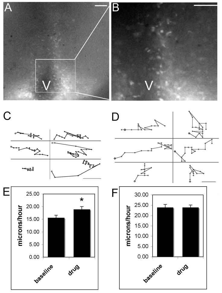
The ventromedial (VMN) and arcuate (ARC) nuclei of the hypothalamus are bilateral nuclear groups at the base of the hypothalamus that are organized through the aggregation of neurons born along the third ventricle that migrate laterally. During development, GABAergic neurons and fibers surround the forming (or primordial) VMN while neurons containing GABA receptors are found within the boundaries of the emerging nucleus. To investigate the role that GABAB receptors play in establishing the VMN, Thy-1 yellow fluorescent protein (YFP) mice were utilized for live video microscopy studies. The Thy-1 promoter drives YFP expression in regions of the hypothalamus during development. Administration of the GABAB receptor antagonist saclofen and the GABAA receptor antagonist bicuculline selectively increased the rate of VMN cell movement in slices placed in vitro at embryonic day 14, when cells that form both the ARC and VMN are migrating away from the proliferative zone surrounding the third ventricle. To further test the role of GABAB receptors in VMN development, GABAB receptor knockout mice were used to examine changes in the positions of phenotypically identified cells within the VMN. Cells containing immunoreactive estrogen receptors (ER) alpha were located in the ventrolateral quadrant of the wild type VMN. In GABABR1 knockout mice, these ERalpha positive neurons were located in more dorsal positions at postnatal day (P) 0 and P4. We conclude that GABA alters cell migration and its effect on final cell positioning may lead to changes in the circuitry and connections within specific nuclei of the developing hypothalamus.


Figures

Figure 1
Figure 1
Brain slices 250μm thick were used for the collection of video microscopy data indicating an increase in neuronal movement in the region of the developing VMN but not the ARC in response to GABA antagonists. The entire region of the hypothalamus that includes the VMN and ARC (A) has fluorescently labeled cells located near the ventricles (V). The boxed region represents the region magnified in (B; scale bars = 100μm). Migrating neurons did not follow strictly radial or dorsal/ ventral movement patterns, rather they changed directions and speeds numerous times within any given time period. Scatter plots for VMN (C) and ARC (D) cells represent sample neurons tracked over 90 minute time periods (scale bars = 5μm) with baseline shown on the left and drug response on the right of each panel. Movement did not occur between every frame and some cells appear to have moved more or farther than others. In the VMN (C,E) the baseline speeds were significantly slower than those observed following drug treatment (10μM bicuculline or 10μM-100μM saclofen; data combined and labeled as drug due to both drugs eliciting the same response). There was no change in the average rate of motion for neurons in the region of the ARC following GABAergic manipulation (F). The scales are different between panels E and F and ARC neurons moved notably faster than those in the region of the VMN.
Figure 2
Figure 2
Dual label images of Thy-1 driven immunoreactive YFP in neurons and GABA receptor subunits. Immunoreactive GABAB receptor subunit R1 (A), and GABAA receptor subunits α1 (D) and β3 (G) were found in both VMN and ARC (not shown) at E15. YFP containing neurons were surrounded by networks of GABA receptor subunit expression (C, F, I). White arrows indicate example cells that appear to show colocalization. For GABABR1 and GABAAβ3 subunits, immunoreactivity within the VMH was apparent in virtually all cells in non-nuclear compartment(s) whereas immunoreactive GABAAα1 subunits were found in a subset of cells (also in non-nuclear cellular compartments). Images were taken on a confocal microscope with a 1.6μm optical slice (scale bars = 50μm).
Figure 3
Figure 3
Images show immunoreactive ERα in sections through the center of the VMN examined at 3 different developmental ages (E17, P0, and P4) in wild-type (A, C, E) and GABABR1 knockout (B, D, F) mice revealed a significant difference in the distribution of ERα positive cells in the ventrolateral quadrant of the VMN. At E17 there was no difference in the position of ERα immunoreactive cells (A and B). At P0 (C and D) and P4 (E and F) there was a more dorsal shift of cells containing immunoreactive ERα in the knockout animal. There was no significant change in ERα expression in the ARC.
Figure 4
Figure 4
Graphic representation of the area of ERα immunoreactivity in GABABR1 wild-type and knockout mice at 3 developmental ages. Bar graphs illustrate the immunoreactivity seen in each row based on a grid system with the first row along the base of the brain (n=6 for each genotype at each age). At P0 (B), the peak of immunoreactivity in the wild-type animal is between 150 and 200μm from the base of the brain and the peak of immunoreactivity in the KO animal is between 250 and 300μm from the base of the brain (scale bars = 100μm). At P4 (C), the peak of immunoreactivity in the wild-type is around 250μm from the base of the brain while the peak in immunoreactivity for the knockout animal ranged from 300-400μm from the base of the brain. There were significant differences between the wild-type and knockout animals at these two ages (genotype × position interaction, p<0.05). There were no significant differences at E17 (A).
Figure 5
Figure 5
BDNF and SF-1 expression in the VMN did not change in the GABABR1 knockout mouse at P0 (scale bars = 100μm). Wild-type (A, D) and GABABR1 knockout (B, E) mice were examined for changes in expression of BDNF mRNA (A-C) and immunoreactive SF-1 protein (D-F) located in the central and dorsomedial regions of the VMN. There were no apparent changes in total expression levels or the distribution of cells containing either marker.
Figure 6
Figure 6
Dual label images show immunoreactive Thy1/YFP (green) and RC2 (red) in the GABABR1 wild-type (A) and knockout (B) mouse. Radial fibers were intact in the knockout mouse suggesting that altered neuronal movement seen in Thy1/YFP-positive neurons was not due to severed or misdirected radial fibers (scale bars = 100μm; standard epifluorescence microscopy). GABABR1 (C) and ERα (D) appear to localize in the same cells (example at white arrow). As GABABR1 is a receptor found on the cell surface and ERα is a nuclear receptor it is difficult to conclude definitive colocalization. Images C, D, and E were taken using a confocal microscope with an optical slice of 1.6μm.
Figure 7
Figure 7
Images show a subset of neurons co-localize immunoreactive Thy-1/YFP with islet-1 or ERα. The ventrolateral VMN contains a small population of ERα immunoreactive neurons at E15 (A) while the ARC contains a larger population of ERα cells (D). For the most part these cells co-localize (white arrows) in only a small number Thy/YFP neurons in the VMN (C) and ARC (F). Islet-1 is also expressed at developmental ages in the ventrolateral VMN and ARC. At E17 there was a subset of cells containing immunoreactive islet-1 (G) that were also Thy-1/YFP-positive (H, I). White arrows indicate examples of cells that colabel with YFP (scale bars = 50μm).
Figure 8
Figure 8
In “normal” VMN development (A) cells migrate laterally, away from the third ventricle. Based upon the cell phenotype, VMN neurons occupy different subdivisions of the nucleus. As illustrated, SF-1 containing neurons occupy the dorsal and central regions of the nucleus while ERα containing neurons occupy the ventrolateral quadrant. Most cell movement throughout the nucleus is radial but there is some dorsal ventral movement, therefore ERα neurons may move in both directions before ending up in their final positions. Dotted arrows indicate movement speed of neurons migrating away from the third ventricle. Based on video microscopy data we know that VMN neurons moving away from the third ventricle move at an average rate of 15μm/hour. When SF-1 signaling is absent there is a medial shift in the positioning of ERα immunoreactive cells coincident with a medial shift of GABAergic neurons and fibers (Dellovade et al., 2000). Panels C and D illustrate changes when GABA signaling is inhibited. When GABAA signaling is reduced (C) there is an increase in the overall size of the VMN (Dellovade et al., 2001), ERα immunoreactivity in the ventrolateral quadrant is spread out and shifted slightly more dorsal. Based on data in the current study a lack of GABAB receptor signaling (D) does not change SF-1 expression throughout the dorsal and central VMN but does result in changes in the ventrolateral ERα population.

Similar articles

Cited by

References

    1. Behar TN, Li YX, Tran HT, Ma W, Dunlap V, Scott C, Barker JL. GABA stimulates chemotaxis and chemokinesis of embryonic cortical neurons via calcium-dependent mechanisms. J Neurosci. 1996;16:1808–1818. - PMC - PubMed
    1. Behar TN, Schaffner AE, Scott CA, O’Connell C, Barker JL. Differential response of cortical plate and ventricular zone cells to GABA as a migration stimulus. J Neurosci. 1998;18:6378–6387. - PMC - PubMed
    1. Berrendero F, Garcia-Gil L, Hernandez ML, Romero J, Cebeira M, de Miguel R, Ramos JA, Fernandez-Ruiz JJ. Localization of mRNA expression and activation of signal transduction mechanisms for cannabinoid receptor in rat brain during fetal development. Development. 1998;125:3179–3188. - PubMed
    1. Bless EP, Walker HJ, Yu KW, Knoll JG, Moenter SM, Schwarting GA, Tobet SA. Live view of gonadotropin-releasing hormone containing neuron migration. Endocrinology. 2005;146:463–468. - PubMed
    1. Bowery NG, Brown DA. The cloning of GABA(B) receptors. Nature. 1997;386:223–224. - PubMed

Publication types

MeSH terms