

The three-dimensional micro- and nanostructure of the aortic medial lamellar unit measured using 3D confocal and electron microscopy imaging
- PMID: 18248974
- PMCID: PMC2679973
- DOI: 10.1016/j.matbio.2007.10.008
The three-dimensional micro- and nanostructure of the aortic medial lamellar unit measured using 3D confocal and electron microscopy imaging
Abstract
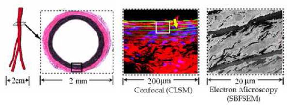
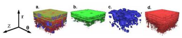
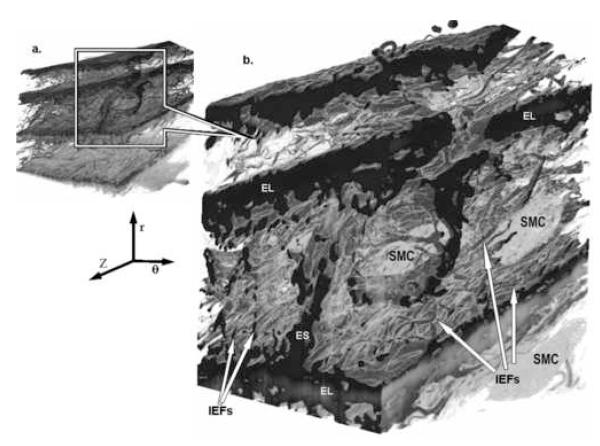
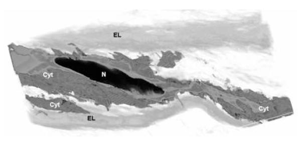
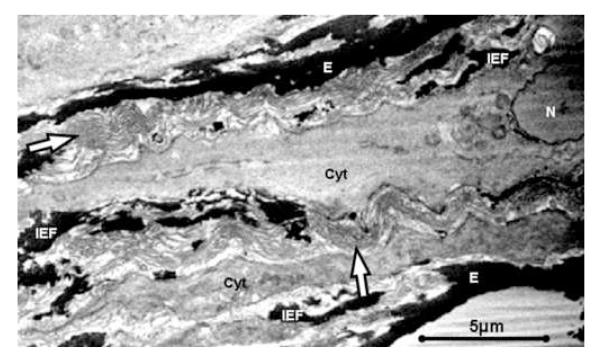
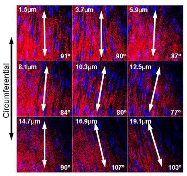
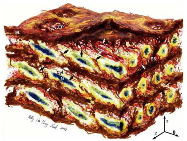
Changes in arterial wall composition and function underlie all forms of vascular disease. The fundamental structural and functional unit of the aortic wall is the medial lamellar unit (MLU). While the basic composition and organization of the MLU is known, three-dimensional (3D) microstructural details are tenuous, due (in part) to lack of three-dimensional data at micro- and nano-scales. We applied novel electron and confocal microscopy techniques to obtain 3D volumetric information of aortic medial microstructure at micro- and nano-scales with all constituents present. For the rat abdominal aorta, we show that medial elastin has three primary forms: with approximately 71% of total elastin as thick, continuous lamellar sheets, 27% as thin, protruding interlamellar elastin fibers (IEFs), and 2% as thick radial struts. Elastin pores are not simply holes in lamellar sheets, but are indented and gusseted openings in lamellae. Smooth muscle cells (SMCs) weave throughout the interlamellar elastin framework, with cytoplasmic extensions abutting IEFs, resulting in approximately 20 degrees radial tilt (relative to the lumen surface) of elliptical SMC nuclei. Collagen fibers are organized as large, parallel bundles tightly enveloping SMC nuclei. Quantification of the orientation of collagen bundles, SMC nuclei, and IEFs reveal that all three primary medial constituents have predominantly circumferential orientation, correlating with reported circumferentially dominant values of physiological stress, collagen fiber recruitment, and tissue stiffness. This high resolution three-dimensional view of the aortic media reveals MLU microstructure details that suggest a highly complex and integrated mural organization that correlates with aortic mechanical properties.
Figures

References
-
- Armentano RL, Levenson J, Barra JG, Fischer EI, Breitbart GJ, Pichel RH, Simon A. Assessment of elastin and collagen contribution to aortic elasticity in conscious dogs. Am J Physiol. 1991;260:H1870–1877. - PubMed
-
- Arner A, Uvelius B. Force-velocity characteristics and active tension in relation to content and orientation of smooth muscle cells in aortas from normotensive and spontaneous hypertensive rats. Circ Res. 1982;50:812–821. - PubMed
-
- Avolio A, Jones D, Tafazzoli-Shadpour M. Quantification of alterations in structure and function of elastin in the arterial media. Hypertension. 1998;32:170–175. - PubMed
-
- Bierring F, Kobayasi T. Electron microscopy of the normal rabbit aorta. Acta Pathol Microbiol Scand. 1963;57:154–168. - PubMed
Publication types
MeSH terms
Substances
Grants and funding
LinkOut - more resources
Full Text Sources
