

doi: 10.1102/1470-7330.2004.0003.
Intensity-modulated radiotherapy--what is it?
Affiliations
- PMID: 18250011
- PMCID: PMC1434586
- DOI: 10.1102/1470-7330.2004.0003
Item in Clipboard
Intensity-modulated radiotherapy--what is it?
Cancer Imaging.
.
Free PMC article
Abstract
Intensity-modulated radiotherapy (IMRT) is one of the most important recent developments in oncology. It enables precise conformation of the radiation dose to the target volume. It has the potential to significantly reduce long-term morbidity and improve local control. This article explains the basic principles of IMRT in comparison to other planning techniques. The current clinical data are presented and future lines of research are discussed.
Figures
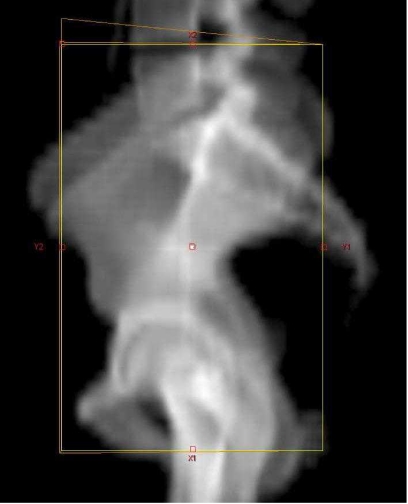
Conventional field (yellow) defined by bony landmarks.
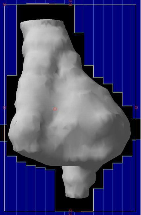
Conformal field. The volume is outlined on each CT slice and a three-dimensional volume is created (white). Fields are shaped with MLC leaves (blue).
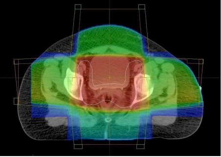
Conformal dose colour-wash. The target volume is contoured in white. The high dose region (red) is brick-shaped and includes part of the bladder.
IMRT fluence represented with colour. The field is divided into multiple beamlets with variable intensities.
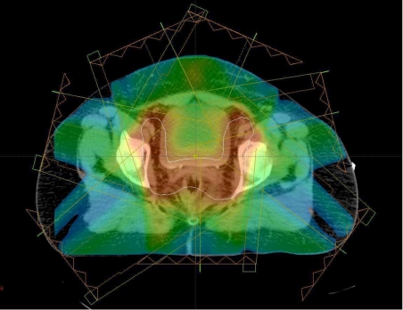
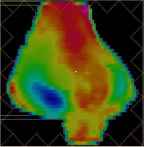
IMRT dose colour-wash. The high dose region (red) conforms to the target volume (white) in a concave shape reducing the bladder and bowel dose.
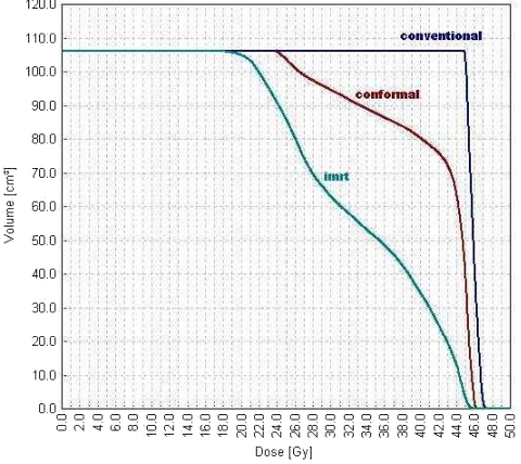
Comparative dose-volume histogram for the three techniques.
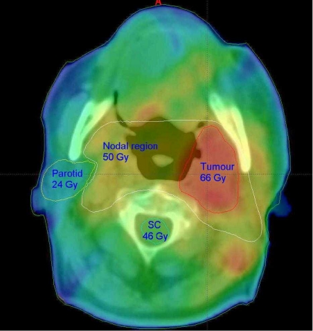
Dose-painting for an oropharyngeal tumour. The target doses for the spinal cord (sc), parotid, lymph nodes and tumour are marked. The IMRT plan delivers a high dose (red) to the tumour and low dose (blue) to the contralateral parotid gland.
References
-
- Dearnaley DP, Khoo VS, Norman AR, et al. Comparison of radiation side-effects of conformal and conventional radiotherapy in prostate cancer: a randomised trial. Lancet. 1999;353(9149):267–72. - PubMed
-
- IMRT Collaborative Working Group Intensity-modulated radiotherapy: current status and issues of interest. Int J Radiat Oncol Biol Phys. 2001;51(4):880–914. - PubMed
-
- Emami B, Sethi A, Petruzzelli GJ. Influence of MRI on target volume delineation and IMRT planning in nasopharyngeal carcinoma. Int J Radiat Oncol Biol Phys. 2003;57(2):481–8. - PubMed
-
- Van Herk M, Bruce A, Kroes AP, Shouman T, Touw A, Lebesque JV. Quantification of organ motion during conformal radiotherapy of the prostate by three dimensional image registration. Int J Radiat Oncol Biol Phys. 1995;33(5):1311–20. - PubMed
-
- Lattanzi J, McNeeley S, Pinover W, et al. A comparison of daily CT localization to a daily ultrasound-based system in prostate cancer. Int J Radiat Oncol Biol Phys. 1999;43(4):719–25. - PubMed
LinkOut - more resources
Full Text Sources