

Defining the tumour and target volumes for radiotherapy
- PMID: 18250025
- PMCID: PMC1434601
- DOI: 10.1102/1470-7330.2004.0054
Defining the tumour and target volumes for radiotherapy
Abstract
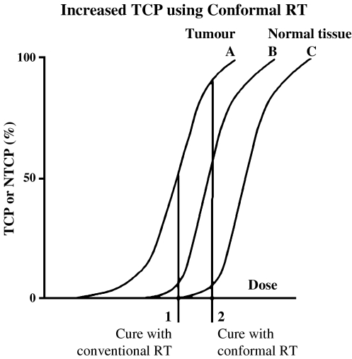
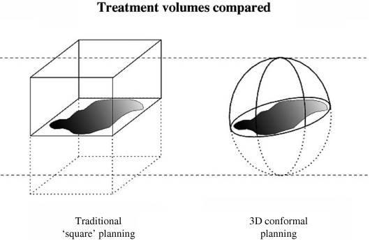
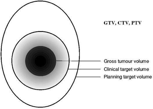
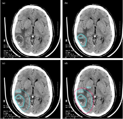
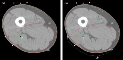
Radiotherapy is a localised treatment. The definition of tumour and target volumes for radiotherapy is vital to its successful execution. This requires the best possible characterisation of the location and extent of tumour. Diagnostic imaging, including help and advice from diagnostic specialists, is therefore essential for radiotherapy planning. There are three main volumes in radiotherapy planning. The first is the position and extent of gross tumour, i.e. what can be seen, palpated or imaged; this is known as the gross tumour volume (GTV). Developments in imaging have contributed to the definition of the GTV. The second volume contains the GTV, plus a margin for sub-clinical disease spread which therefore cannot be fully imaged; this is known as the clinical target volume (CTV). It is the most difficult because it cannot be accurately defined for an individual patient, but future developments in imaging, especially towards the molecular level, should allow more specific delineation of the CTV. The CTV is important because this volume must be adequately treated to achieve cure. The third volume, the planning target volume (PTV), allows for uncertainties in planning or treatment delivery. It is a geometric concept designed to ensure that the radiotherapy dose is actually delivered to the CTV. Radiotherapy planning must always consider critical normal tissue structures, known as organs at risk (ORs). In some specific circumstances, it is necessary to add a margin analogous to the PTV margin around an OR to ensure that the organ cannot receive a higher-than-safe dose; this gives a planning organ at risk volume. This applies to an organ such as the spinal cord, where damage to a small amount of normal tissue would produce a severe clinical manifestation. The concepts of GTV, CTV and PTV have been enormously helpful in developing modern radiotherapy. Attention to detail in radiotherapy planning is vital, and does affect outcomes: 'the devil is in the detail'. Radiotherapy planning is also dependent on high quality imaging, and the better the imaging the better will be the outcomes from radiotherapy.
Figures
Similar articles
-
[Target volume concepts in radiotherapy and their implications for imaging].Radiologe. 2018 Aug;58(8):708-721. doi: 10.1007/s00117-018-0420-6. Radiologe. 2018. PMID: 29951925 Review. German.
-
Three-dimensional dosimetric evaluation of a conventional radiotherapy technique for treatment of nasopharyngeal carcinoma.Radiother Oncol. 2001 Feb;58(2):143-53. doi: 10.1016/s0167-8140(00)00336-4. Radiother Oncol. 2001. PMID: 11166865
-
Is a clinical target volume (CTV) necessary for locally advanced non-small cell lung cancer treated with intensity-modulated radiotherapy? -a dosimetric evaluation of three different treatment plans.J Thorac Dis. 2017 Dec;9(12):5194-5202. doi: 10.21037/jtd.2017.10.147. J Thorac Dis. 2017. PMID: 29312726 Free PMC article.
-
The value of magnetic resonance imaging in target volume delineation of base of tongue tumours--a study using flexible surface coils.Radiother Oncol. 2010 Feb;94(2):161-7. doi: 10.1016/j.radonc.2009.12.021. Epub 2010 Jan 22. Radiother Oncol. 2010. PMID: 20096947
-
The role of computational methods for automating and improving clinical target volume definition.Radiother Oncol. 2020 Dec;153:15-25. doi: 10.1016/j.radonc.2020.10.002. Epub 2020 Oct 8. Radiother Oncol. 2020. PMID: 33039428 Review.
Cited by
-
DTI Abnormalities Related to Glioblastoma: A Prospective Comparative Study with Metastasis and Healthy Subjects.Curr Oncol. 2022 Apr 16;29(4):2823-2834. doi: 10.3390/curroncol29040230. Curr Oncol. 2022. PMID: 35448204 Free PMC article.
-
Advancing the Collaboration Between Imaging and Radiation Oncology.Semin Radiat Oncol. 2024 Oct;34(4):402-417. doi: 10.1016/j.semradonc.2024.07.005. Semin Radiat Oncol. 2024. PMID: 39271275 Review.
-
Circulating Tumor DNA-Based Disease Monitoring of Patients with Locally Advanced Esophageal Cancer.Cancers (Basel). 2022 Sep 11;14(18):4417. doi: 10.3390/cancers14184417. Cancers (Basel). 2022. PMID: 36139577 Free PMC article.
-
Development and prospective in-patient proof-of-concept validation of a surface photogrammetry + CT-based volumetric motion model for lung radiotherapy.Med Phys. 2019 Dec;46(12):5407-5420. doi: 10.1002/mp.13824. Epub 2019 Oct 25. Med Phys. 2019. PMID: 31518437 Free PMC article.
-
Fully automated segmentation of clinical target volume in cervical cancer from magnetic resonance imaging with convolutional neural network.J Appl Clin Med Phys. 2022 Sep;23(9):e13725. doi: 10.1002/acm2.13725. Epub 2022 Jul 27. J Appl Clin Med Phys. 2022. PMID: 35894782 Free PMC article.
References
-
- Swedish Council on Technology Assessment in Health Care (SBU) Radiotherapy for cancer. Acta Oncol. 1996;35(Suppl 6):1–100. (Suppl 7): 1–152.
-
- Suit H. Contributions of L.H. Gray to radiation physics, biology, and oncology. Int J Radiat Oncol Biol Phys. 2002;53(4):795–7. - PubMed
-
- Burnet NG, Wurm R, Nyman J, Peacock JH. Normal tissue radiosensitivity—how important is it? Clin Oncol. 1996;8:25–34. - PubMed
-
- Horiot JC, Le Fur R, N’Guyen T, et al. Hyperfractionation versus conventional fractionation in oropharyngeal carcinoma: final analysis of a randomised trial of the EORTC cooperative group of radiotherapy. Radiother Oncol. 1992;25(4):229–30. - PubMed
-
- Yarnold JR, Owen JR, Ashton A, et al. Fractionation sensitivity of change in breast appearance after radiotherapy for early breast cancer: long-term results of a randomised trial. Radiother Oncol. 2002;64(Suppl 1):S25. - PubMed
LinkOut - more resources
Full Text Sources