. 2008 Feb 18;205(2):385-93.
doi: 10.1084/jem.20071507. Epub 2008 Feb 4.

Ozone exposure in a mouse model induces airway hyperreactivity that requires the presence of natural killer T cells and IL-17

Affiliations

Ozone exposure in a mouse model induces airway hyperreactivity that requires the presence of natural killer T cells and IL-17

Muriel Pichavant et al. J Exp Med. .

Abstract

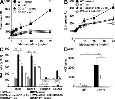
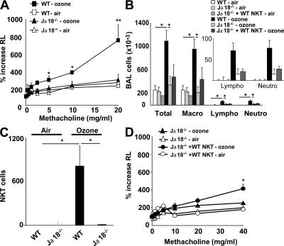
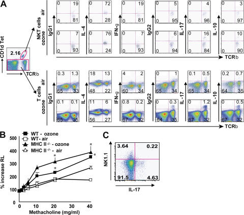
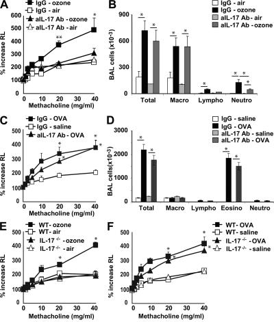
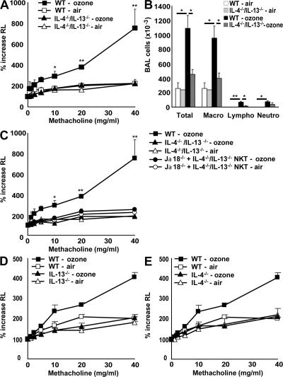
Exposure to ozone, which is a major component of air pollution, induces a form of asthma that occurs in the absence of adaptive immunity. Although ozone-induced asthma is characterized by airway neutrophilia, and not eosinophilia, it is nevertheless associated with airway hyperreactivity (AHR), which is a cardinal feature of asthma. Because AHR induced by allergens requires the presence of natural killer T (NKT) cells, we asked whether ozone-induced AHR had similar requirements. We found that repeated exposure of wild-type (WT) mice to ozone induced severe AHR associated with an increase in airway NKT cells, neutrophils, and macrophages. Surprisingly, NKT cell-deficient (CD1d(-/-) and Jalpha18(-/-)) mice failed to develop ozone-induced AHR. Further, treatment of WT mice with an anti-CD1d mAb blocked NKT cell activation and prevented ozone-induced AHR. Moreover, ozone-induced, but not allergen-induced, AHR was associated with NKT cells producing interleukin (IL)-17, and failed to occur in IL-17(-/-) mice nor in WT mice treated with anti-IL-17 mAb. Thus, ozone exposure induces AHR that requires the presence of NKT cells and IL-17 production. Because NKT cells are required for the development of two very disparate forms of AHR (ozone- and allergen-induced), our results strongly suggest that NKT cells mediate a unifying pathogenic mechanism for several distinct forms of asthma, and represent a unique target for effective asthma therapy.


Figures

Figure 1.
Figure 1.
Ozone exposure induces AHR and increases airway inflammation in WT, but not in CD1d−/−, mice. WT BALB/c mice and NKT cell–deficient CD1d−/− mice were exposed 3 times to 1 ppm of ozone over 5 d or to room air. (A) Changes in lung resistance (RL) were measured in anesthetized, tracheotomized, intubated, and mechanically ventilated mice. Ozone exposure induces an increase in AHR in WT, but not in CD1d−/−, mice. (B) WT BALB/c and CD1d−/− mice were exposed to ozone as in A, and treated with anti-CD1d blocking mAb or isotype control mAb. Ozone-induced AHR was prevented by the anti-CD1d mAb treatment. (C) BAL fluid was collected 24 h after the last ozone challenge of mice depicted in A and B. Total and differential cell counts were evaluated in BAL fluid. Ozone exposure induced pulmonary inflammation associated with neutrophils, which was reduced by treatment with anti-CD1d mAb and in CD1d−/− mice. The inset is a magnification of the data for lymphocytes and neutrophils. (D) Ozone exposure increased the number of iNKT cells in BAL fluid of WT, but not CD1d−/−, mice. Anti-CD1d mAb treatment decreased the number of iNKT cells in BAL fluid after ozone exposure. iNKT cells were identified by staining with CD1d tetramers loaded with PBS57 (or with empty CD1d tetramers as control) and anti-TCRβ mAb. Results are expressed as the mean ± the SEM. P ≤ 0.05 (*) and P ≤ 0.01 (**) compared with mice exposed to air. These results represent one out of four experiments, with five mice in each group.
Figure 2.
Figure 2.
Ozone exposure induces AHR in WT, but not in Jα18−/−, mice. WT BALB/c and iNKT cell–deficient Jα18−/− mice were exposed 3 times to 1 ppm of ozone versus air. (A) Changes in lung resistance (RL) were measured on anesthetized, tracheotomized, intubated, and mechanically ventilated mice. Ozone exposure induced significant AHR in WT, but not in Jα18−/− mice. (B) Total and differential cell counts in BAL fluid were evaluated. WT, but not Jα18−/−, mice develop an increase in the number of airway macrophages, lymphocytes, and neutrophils. The insert is a magnification of the data for lymphocytes and neutrophils. (C) Ozone exposure increases iNKT cell number in BAL fluid of WT, but not Jα18−/−, mice. NKT cells were identified as described in Fig. 1 D. (D) Adoptive transfer of WT iNKT cells into Jα18−/− mice partially restores AHR induced by repeated ozone exposure. Results are expressed as the mean ± the SEM. P ≤ 0.05 (*) and P ≤ 0.01 (**) compared with mice exposed to air. These results represent one out of four experiments, with five mice in each group.
Figure 3.
Figure 3.
iNKT cells and T cells produce IL-17 after ozone exposure. WT BALB/c mice were exposed 3 times to 1 ppm of ozone versus air. Intracellular IL-4, IFN-γ, IL-17, and IL-10 staining was performed on T cell–enriched lung cells without further stimulation, but treated with GolgiStop for 2 h. (A) iNKT cells were analyzed by gating on CD1d tetramer+ TCRβ+ cells (top). T cells were analyzed by gating on CD1d tetramer cells (bottom). The flow cytometry data are provided as dot plots. Numbers in each quadrant indicate the percentage of cells in that quadrant. The number of dots (events) is much greater for T cells than for iNKT cells because the number of NKT cells in the lungs is only a fraction of the number of T cells present. Data are representative of three independent experiments. (B) MHC class II−/− mice were exposed to ozone or air. Changes in lung resistance (RL) were measured on anesthetized, tracheotomized, intubated, and mechanically ventilated mice. Ozone exposure induced significant AHR in WT mice and MHC class II−/− mice. (C) C57BL/6 mice were exposed to ozone. iNKT cells in the BAL fluid were analyzed by gating on CD1d tetramer+ TCRβ+ cells, and by staining for expression of NK1.1 and IL-17. The IL-17+ cells were NK1.1 negative.
Figure 4.
Figure 4.
IL-17 is involved in ozone-induced, but not in OVA-induced, AHR. (A and B) The response of WT mice to ozone was greatly reduced by treatment with anti–IL-17 mAb (aIL-17 Ab). WT mice were exposed 3 times to 1 ppm ozone versus air and treated with anti–IL-17 blocking mAb or isotype control. (A) Airway responsiveness to methacholine was measured in anesthetized, tracheotomized, intubated, and mechanically ventilated mice and reported as the percentage of increase in airway resistance. (B) BAL fluid was collected 24 h after the last airway challenge. Total and differential cell counts were evaluated. Treatment with anti–IL-17 reduced airway lymphocytes and neutrophils. (C and D) Anti–IL-17 Ab treatment does not reduce OVA-induced AHR (C) or OVA-induced airway inflammation (D). Mice were sensitized i.p. and challenged intranasally with OVA for 3 d, and were treated with anti–IL-17 blocking mAb or isotype control 1 d before the 3 OVA challenges. (D) BAL fluid was collected and evaluated as in B. Results are expressed as the mean ± the SEM. P ≤ 0.05 (*) and P ≤ 0.01 (**) compared with untreated mice or mice exposed to air. Data are representative of two independent experiments with five mice in each group. (E) IL-17−/− mice fail to develop ozone-induced AHR. Mice were treated as in A and B, and examined for AHR. (F) IL-17−/− mice develop normal OVA-induced AHR. IL-17−/− mice were sensitized and challenged with OVA before examination for AHR by challenge with methacholine.
Figure 5.
Figure 5.
IL-4 and -13 are involved in ozone-induced AHR and airway inflammation. WT BALB/c, IL-4−/−/IL-13−/− double-knockout, IL-13−/−, and IL-4−/− mice were exposed 3 times to 1 ppm of ozone versus air. (A) AHR was measured as in Fig. 4 A. IL-4−/−/IL-13−/− double-knockout mice failed to develop ozone-induced AHR. (B) Total and differential cell counts were evaluated in BAL fluid. IL-4−/−/IL-13−/− double-knockout mice failed to develop ozone-induced airway inflammation. (C) Adoptive transfer of IL-4−/−/IL-13−/− iNKT cells into Jα18−/− failed to restore ozone-induced AHR. Results are expressed as the mean ± the SEM. P ≤ 0.05 (*) and P ≤ 0.01 (**) compared with mice exposed to air, and double-knockout mice. These results are representative of three experiments. (D) IL-13−/− mice were exposed to ozone before measurement of AHR, as in A. AHR failed to occur in the IL-13−/− mice. (E) IL-4−/− mice were exposed to ozone before measurement of AHR, as in A. AHR failed to occur in the IL-4−/− mice.

References

    1. Gold, D.R., and R. Wright. 2005. Population disparities in asthma. Annu. Rev. Public Health. 26:89–113. - PubMed
    1. Wills-Karp, M. 1999. Immunologic basis of antigen-induced airway hyperresponsiveness. Annu. Rev. Immunol. 17:255–281. - PubMed
    1. Voehringer, D., T.A. Reese, X. Huang, K. Shinkai, and R.M. Locksley. 2006. Type 2 immunity is controlled by IL-4/IL-13 expression in hematopoietic non-eosinophil cells of the innate immune system. J. Exp. Med. 203:1435–1446. - PMC - PubMed
    1. Gibson, P.G., J.L. Simpson, and N. Saltos. 2001. Heterogeneity of airway inflammation in persistent asthma: evidence of neutrophilic inflammation and increased sputum interleukin-8. Chest. 119:1329–1336. - PubMed
    1. Li, N., J. Alam, M.I. Venkatesan, A. Eiguren-Fernandez, D. Schmitz, E. Di Stefano, N. Slaughter, E. Killeen, X. Wang, A. Huang, et al. 2004. Nrf 2 is a key transcription factor that regulates antioxidant defense in macrophages and epithelial cells: protecting against the proinflammatory and oxidizing effects of diesel exhaust chemicals. J. Immunol. 173:3467–3481. - PubMed

Publication types

MeSH terms