. 2008 Feb 12;105(6):2169-74.
doi: 10.1073/pnas.0711647105. Epub 2008 Feb 4.

Bcl-xL induces Drp1-dependent synapse formation in cultured hippocampal neurons

Affiliations

Bcl-xL induces Drp1-dependent synapse formation in cultured hippocampal neurons

Hongmei Li et al. Proc Natl Acad Sci U S A. .

Abstract

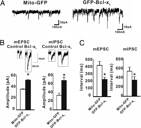
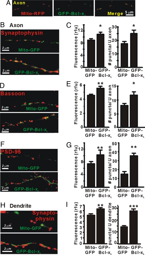
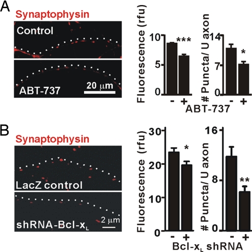
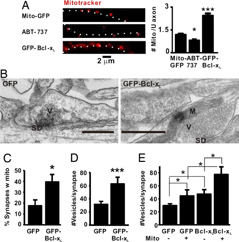
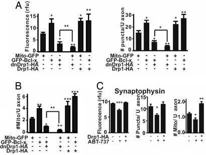
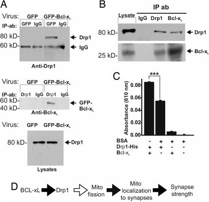
Maturation of neuronal synapses is thought to involve mitochondria. Bcl-xL protein inhibits mitochondria-mediated apoptosis but may have other functions in healthy adult neurons in which Bcl-xL is abundant. Here, we report that overexpression of Bcl-xL postsynaptically increases frequency and amplitude of spontaneous miniature synaptic currents in rat hippocampal neurons in culture. Bcl-xL, overexpressed either pre or postsynaptically, increases synapse number, the number and size of synaptic vesicle clusters, and mitochondrial localization to vesicle clusters and synapses, likely accounting for the changes in miniature synaptic currents. Conversely, knockdown of Bcl-xL or inhibiting it with ABT-737 decreases these morphological parameters. The mitochondrial fission protein, dynamin-related protein 1 (Drp1), is a GTPase known to localize to synapses and affect synaptic function and structure. The effects of Bcl-xL appear mediated through Drp1 because overexpression of Drp1 increases synaptic markers, and overexpression of the dominant-negative dnDrp1-K38A decreases them. Furthermore, Bcl-xL coimmunoprecipitates with Drp1 in tissue lysates, and in a recombinant system, Bcl-xL protein stimulates GTPase activity of Drp1. These findings suggest that Bcl-xL positively regulates Drp1 to alter mitochondrial function in a manner that stimulates synapse formation.


Conflict of interest statement

The authors declare no conflict of interest.

Figures

Fig. 1.
Fig. 1.
Bcl-xL alters the spontaneous release of neurotransmitter. (A) Representative recordings at −70 mV of spontaneous mEPSCs and mIPSCs of rat hippocampal neurons transfected (DIV5) with Mito-GFP (control) or GFP-Bcl-xL. (B) Recordings and quantified amplitudes of mEPSCs and mIPSCs for all experiments, as in A (DIV17) (n = 6 cells for GFP-Bcl-xL; n = 8 for Mito-GFP). (C) Quantified intervals between events for all experiments. *, P < 0.05, determined by Student's t test.
Fig. 2.
Fig. 2.
Bcl-xL induces synapse formation in cultured hippocampal neurons. (A) Fluorescence microscopy of an axon (DIV13) of a cultured hippocampal neuron cotransfected (DIV5) with Mito-RFP (red) and GFP-Bcl-xL (green) and the merge of the two images. (B and C) Immunofluorescence microscopy for synaptophysin in axons of transfected neurons. Fluorescence intensity of puncta in relative fluorescence units (rfu) and their number per unit length (≈35 μm) of axon are presented as mean ± SEM for n = 15 neurons per group in three independent experiments. *, P < 0.05; **, P < 0.01, Student's t test. (D and E) Immunofluorescence microscopy for bassoon as in B and C. (F and G) Immunofluorescence microscopy for PSD-95 as in B and C. (H and I) Immunofluorescence microscopy for synaptophysin puncta apposed to dendrites of transfected neurons as in B and C. Synapse formation is highly likely in H, but dendrites may or may not be present in B, D, and F.
Fig. 3.
Fig. 3.
Bcl-xL is required for synapse formation. (A) Immunofluorescence microscopy for synaptophysin in axons of untransfected hippocampal neurons (DIV14) with or without 1 μM ABT-737 added at DIV5. Data were quantified as in Fig. 2C for three independent experiments (n = 6 cells per group). *, P < 0.05; ***, P < 0.001, Student's t test. White dots indicate the course of the axon. (B) Immunofluorescence microscopy for synaptophysin in axons of hippocampal neurons (DIV14) that were transfected (DIV5) with bcl-x hairpin RNA (pCAG-lacZ-shRNA-Bcl-xL) or control vector (pCAG-lacZ). Synaptophysin intensity (rfu) and number of puncta per 20 μm of axon in three independent experiments were quantified (mean ± SEM; n = 12 neurons per group). *, P < 0.05; **, P < 0.01; ***, P < 0.001, Student's t test.
Fig. 4.
Fig. 4.
Bcl-xL increases the number of mitochondria per unit length of axon and their colocalization to presynaptic sites. (A) Fluorescence microscopy of transfected axons in hippocampal cultures stained with MitoTracker Red. Neurons were either transfected as indicated or treated with ABT-737 (DIV5 for both). White dots beside the axon images show the axon trajectories. The number of MitoTracker-stained mitochondria per 6 μm of axon (mean ± SEM) was quantified for both control and ABT-737 (n = 6 neurons per group) and for GFP-Bcl-xL (n = 5 in each of three independent experiments for a total of 15 neurons). *, P < 0.05; ***, P < 0.001. (B) Representative electron micrographs of synaptic profiles identified by a synaptic density between neurons (DIV13) expressing GFP only or GFP-Bcl-xL. Data are quantified in C–E. V, vesicles; SD, synaptic density; M, mitochondrion. (Scale bars: 500 nm.) (C) Percentage of synaptic profiles containing a mitochondrion. (D) Number of vesicles per synaptic profile. (E) The number of vesicles per synaptic profile was greater if a mitochondrion was present and if the neuron expressed GFP-BclxL. *, P < 0.05; ***, P < 0.001.
Fig. 5.
Fig. 5.
Drp1-dependent function of Bcl-xL in synapse formation and mitochondrial localization. (A) Quantified immunofluorescence intensity (rfu, Left) and number of synaptophysin puncta per 20 μm of axon (mean ± SEM, Right) for three independent experiments as in Fig. 2 (n = total of 12 neurons per group). *, P < 0.05; **, P < 0.01 compared with Mito-GFP control, Student's t test. Significance of difference between Mito-GFP and GFP-Bcl-xL each with dnDrp1-K38A is also indicated. (B) Quantified number of mitochondria per 6 μm of axon in three independent experiments (mean ± SEM; n = 10 neurons per group). *, P < 0.05; **, P < 0.01; ***, P < 0.001, Student's t test and comparisons as in A. (C) Quantified immunofluorescence for synaptophysin intensity (rfu) and number of puncta per 20 μm of axon and number of mitochondria per 6 μm of axon of hippocampal neurons (DIV13) transfected with Drp1-HA (DIV5) and/or treated with ABT-737 (DIV5) (means ± SEM, n = 6 cells per group in each of three independent experiments). *, P < 0.05; **, P < 0.01; ***, P < 0.001 compared with control.
Fig. 6.
Fig. 6.
Bcl-xL and Drp1 interact. (A) (Top and Middle) Immunoblots of reciprocal coimmunoprecipitation experiments on hippocampal neurons (DIV13) transduced with viral vectors (DIV5) as indicated (n = 2). Antibodies used for precipitation (IP) and for immunoblots are indicated. (Top) Drp1 was coprecipitated with GFP-Bcl-xL, brought down by a GFP antibody. (Bottom) Blot of endogenous Drp1 in the starting lysates for immunoprecipitations above. (B) Immunoblots for endogenous Bcl-xL and endogenous Drp1 from adult rat brain after immunoprecipitation with the antibodies indicated. Each panel shows the starting lysate, control IgG precipitate, and the indicated precipitates probed with Drp1 and Bcl-xL antibodies. (C) Relative amounts of phosphate produced by GTPase activity in 1 h with purified recombinant proteins. Shown are results of three independent experiments (mean ± SEM). BSA had no GTPase activity above background, and this signal was subtracted from the other values. ***, P < 0.001. (D) Model of Bcl-xL-induced synaptic function. Black arrows indicate steps addressed in this work.

References

    1. Sanes JR, Lichtman JW. Development of the vertebrate neuromuscular junction. Annu Rev Neurosci. 1999;22:389–442. - PubMed
    1. Young SH, Poo MM. Spontaneous release of transmitter from growth cones of embryonic neurones. Nature. 1983;305:634–637. - PubMed
    1. Chow I, Poo MM. Release of acetylcholine from embryonic neurons upon contact with muscle cell. J Neurosci. 1985;5:1076–1082. - PMC - PubMed
    1. Zhang W, Benson DL. Development and molecular organization of dendritic spines and their synapses. Hippocampus. 2000;10:512–526. - PubMed
    1. Ziv NE, Garner CC. Cellular and molecular mechanisms of presynaptic assembly. Nat Rev Neurosci. 2004;5:385–399. - PubMed

Publication types