. 2008 Feb 12;105(6):2214-9.
doi: 10.1073/pnas.0711453105. Epub 2008 Feb 4.

Resetting of FLOWERING LOCUS C expression after epigenetic repression by vernalization

Affiliations

Resetting of FLOWERING LOCUS C expression after epigenetic repression by vernalization

Candice C Sheldon et al. Proc Natl Acad Sci U S A. .

Abstract

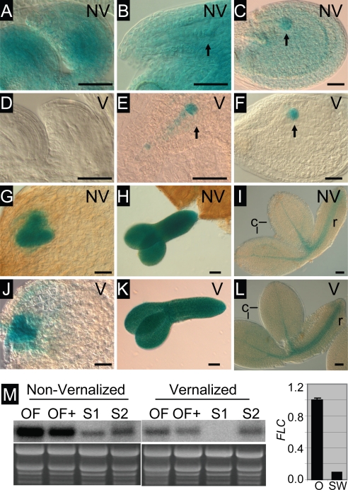
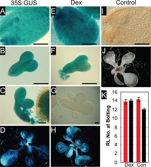
The epigenetic repression of FLOWERING LOCUS C (FLC) in winter-annual ecotypes of Arabidopsis by prolonged cold ensures that plants flower in spring and not during winter. Resetting of the FLC expression level in progeny is an important step in the life cycle of the plant. We show that both the paternally derived and the maternally derived FLC:GUS genes are reset to activity but that the timing of their first expression differs. The paternal FLC:GUS gene in vernalized plants is expressed in the male reproductive organs, the anthers, in both somatic tissue and in the sporogenous pollen mother cells, but there is no expression in mature pollen. In the progeny generation, the paternally derived FLC:GUS gene is expressed in the single-celled zygote (fertilized egg cell) and through embryo development, but not in the fertilized central cell, which generates the endosperm of the progeny seed. FLC:GUS is not expressed during female gametogenesis, with the maternally derived FLC:GUS being first expressed in the early multicellular embryo. We show that FLC activity during late embryo development is a prerequisite for the repressive action of FLC on flowering.


Conflict of interest statement

The authors declare no conflict of interest.

Figures

Fig. 1.
Fig. 1.
FLC-C24:GUS is reset in the somatic and sporogenous tissues in the anther. (A and B) GUS-stained buds from nonvernalized (A) and vernalized (B) C24 + FLC-C24:GUS plants. (C) Quantitative RT-PCR comparing FLC expression in nonvernalized (NV) and vernalized (V) unopened buds and comparing expression in stamens from young buds (S) and remaining bud tissue (B), indicating that expression in young vernalized buds is largely limited to stamens. (D–M) Transverse sections through consecutive buds from vernalized C24 + FLC-C24:GUS plants viewed under dark-field conditions in which the GUS crystals appear pink (F and G are higher-magnification images of D and E, respectively). (D and F) Flower bud at late stage 8, anther stage 4 with sporogenous cells. (E and G) Flower bud at early stage 9, anthers at stage 5 with premeiotic or early meiotic PMC. (H) Stage-6 anther with PMC in meiosis. (I) Stage-7 anther containing tetrads. (J and K) Stage-8 anther with haploid microspores showing induction of expression in tapetum. (L) Stage-10 to -11 anther with degenerating tapetum. (M) Stage-12 anther. The tapetum has degenerated, and two pollen mitoses are complete. a, anther; m, microspore; p, pistil; s, sporogenous cells; t, tapetum; te, tetrad. (Scale bars: 50 μm.) Flower stages are from ref. , and anther stages are from ref. .
Fig. 2.
Fig. 2.
FLC:GUS is not expressed during female gametogenesis, and the paternally derived gene is expressed in the single-celled zygotes from vernalized plants. (A and D) Youngest open pollinated flower of nonvernalized Ler FRI + FLC-Col:GUS. (B and E) Youngest open pollinated flower of vernalized Ler FRI + FLC-Col:GUS. (C and F) Second youngest open pollinated flower of vernalized Ler FRI + FLC-Col:GUS. (A–C) GUS-stained viewed with DIC optics. (D–F) Cleared ovules for stage comparison. (G and J) Sections of C24 + FLC-C24:GUS through ovule primordia around the time of meiosis (G) and at the functional megaspore stage (J) viewed under dark-field conditions. (H and K) Ovules resulting from a cross between male Ler FRI + FLC-Col:GUS vernalized and female Ler FRI-Sf2 vernalized. (I and L) Ovules resulting from a cross between female Ler FRI + FLC-Col:GUS vernalized and male Ler FRI-Sf2 vernalized. (H and I) Single-celled zygote stage ovules (1 day after pollination). (K and L) Early-globular stage embryos (3 days after pollination). Black arrows, endosperm nuclei; red arrows, zygotic nuclei. (Scale bars: 50 μm.)
Fig. 3.
Fig. 3.
FLC-C24:GUS is expressed throughout embryo development. (A–L) GUS-stained ovules and embryos from nonvernalized (NV) or vernalized (V) C24 + FLC-C24:GUS plants. (A and D) Unfertilized ovules. (B and E) Ovules with single-cell pro-embryo and suspensor indicated by arrow. (C and F) Ovules with globular embryo indicated by arrow. (G and J) Ovules with heart-stage embryo. (H and K) Torpedo-stage embryos. (I and L) Late-stage (bent cotyledon) embryos. (M Left) RNA gel blot showing FLC expression in flowers and siliques from nonvernalized and vernalized C24 plants. OF, open flowers; OF+, open flowers with up to 5-mm siliques (containing pro-embryos up to the four-cell stage); S1, siliques between 5 and 10 mm in length (containing embryos from four-cell to globular stage); S2, siliques >10 mm (containing embryos larger than globular stage). The ethidium bromide-stained gel is shown as a loading control. (M Right) Quantitative RT-PCR comparing FLC expression in ovules (O) and silique walls (SW) from ≈10-mm siliques from vernalized plants.
Fig. 4.
Fig. 4.
FLC expression in late embryogenesis is required for delayed flowering. (A–D) 35S:GUS as a marker for 35S:FLC. (E–H) Dexamethasone-treated LhGR/pOp-FLC/GUS in Ler. (I and J) Control-treated LhGR/pOp-FLC/GUS in Ler. (Scale bars: 100 μm.) (K) Flowering time measurements of LhGR/pOp-FLC/GUS in Ler, either dexamethasone-treated (Dex) or control-treated (Con). Seeds for the flowering time experiment came from plants that were either dexamethasone-treated (red bars) or control-treated (black bars) throughout vegetative growth, flowering, and seed development. Error bars indicate the standard error.
Fig. 5.
Fig. 5.
FLC expression under the control of heterologous promoters results in delayed flowering. (A) Flowering time measurement of F1 plants of promoter:LhG4 × LhGh/pOp-FLC/GUS in Ler (red bars) and the corresponding promoter:LhG4 line in Ler (black bars), with the promoter indicated on the x axis. The standard errors are indicated. (B, E, H, K, and N) Ovules containing heart-stage embryos. (C, F, I, L, and O) Dissected bent-cotyledon-stage embryos. (D, G, J, M, and P) Ten-day-old vegetative plants. The promoter driving FLC and GUS expression is indicated at the top. (Scale bars: 100 μm.)

References

    1. Hepworth SR, Valverde F, Ravenscroft D, Mouradov A, Coupland G. Antagonistic regulation of flowering-time gene SOC1 by CONSTANS and FLC via separate promoter motifs. EMBO J. 2002;21:4327–4337. - PMC - PubMed
    1. Helliwell CA, Wood CC, Robertson M, Peacock WJ, Dennis ES. The Arabidopsis FLC protein interacts directly in vivo with SOC1 and FT chromatin and is part of a high-molecular-weight protein complex. Plant J. 2006;46:183–192. - PubMed
    1. Searle I, et al. The transcription factor FLC confers a flowering response to vernalization by repressing meristem competence and systemic signalling in Arabidopsis. Genes Dev. 2006;20:898–912. - PMC - PubMed
    1. Sheldon CC, et al. The FLF MADS box gene: A repressor of flowering in Arabidopsis regulated by vernalization and methylation. Plant Cell. 1999;11:445–458. - PMC - PubMed
    1. Michaels SD, Amasino RM. FLOWERING LOCUS C encodes a novel MADS domain protein that acts as a repressor of flowering. Plant Cell. 1999;11:949–956. - PMC - PubMed

MeSH terms

Substances