Comparative Study
. 2008 Feb 15;180(4):2573-80.
doi: 10.4049/jimmunol.180.4.2573.

A CD8+/CD103high T cell subset regulates TNF-mediated chronic murine ileitis

Affiliations
Comparative Study

A CD8+/CD103high T cell subset regulates TNF-mediated chronic murine ileitis

Johnson Ho et al. J Immunol. .

Abstract

Recruitment of lymphocytes to sites of inflammation requires the sequential engagement of adhesion molecules and chemokine receptors. Of these, the lectin-like molecule CD44 has been particularly implicated in inflammatory trafficking. Using a TNF-driven model of chronic ileitis (i.e., B6.129P-Tnf(Delta)(ARE) mice) that recapitulates many features of Crohn's disease, we demonstrate dynamic changes in the expression and functional state of CD44 on CD8+ T cells. These cells coexpress CD44 and L-selectin, giving them a surface phenotype similar to that of central memory T cells. Yet functionally they exhibit the phenotype of effector T cells, because they produce IFN-gamma. Unexpectedly, depletion of the CD8+ population had no effect on the severity of ileitis. Further analyses showed a second CD8+ population that lacked CD44, but expressed CD103, produced TGF-beta, inhibited the proliferation of CD4+ in vitro, and attenuated adoptively transferred ileitis in vivo, most likely counteracting the proinflammatory role of the CD44(high) subset. Collectively, these data suggest that the presence or absence of CD44 and CD103 on the CD8+ lymphocyte surface defines functionally distinct subsets of CD8+ T cells in vivo. These inflammation-driven populations exert distinct roles during the development of chronic ileitis, and influence the balance of effector and regulatory functions in the chronically inflamed small intestine.


Figures

FIGURE 1
FIGURE 1
Expansion of the CD8+/CD44high subset in TNFΔARE (ΔARE) mice compared with noninflamed age-matched WT littermates. Lymphocytes isolated from the indicated lymphoid compartments at the indicated ages were incubated with anti-CD8+ and allophycocyanin-labeled anti-CD44 mAbs and analyzed by flow cytometry using CD44-deficient lymphocytes (dotted line) from the respective organs and isotype Ab (MFI < 101; data not shown) as controls. Cells were gated on forward scatter (FSC), side scatter (SSC), and CD8. Representative histograms were obtained from four to six mice per group at 4 and ≥20 wk of age.
FIGURE 2
FIGURE 2
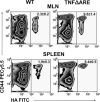
Enhanced hyaluronate binding by CD8+ T cells from TNFΔARE mice compared with noninflamed WT littermates. Freshly isolated CD8+ T cells from the indicated organs were incubated with FITC-labeled hyaluronate (HA FITC) at 37°C for 30 min and analyzed by flow cytometry. Preincubation with nonfluorescent hyaluronate was used to test the specificity of binding (data not shown). Cells were gated on FSC, SSC, and CD8+. Representative density plots were obtained from three experiments using cells from four mice per strain at ≥20 wk of age run in triplicate. Mean hemagglutinin binding ± SEM pooled from three experiments, p < 0.01.
FIGURE 3
FIGURE 3
The CD8+/CD44high T cell population coexpresses L-selectin and CD45RB. A and B, Lymphocytes isolated from the indicated lymphoid compartments were incubated with anti-CD8, anti-CD44, anti-CD45RB, and anti-L-selectin mAbs and analyzed by flow cytometry. Representative density plots and mean ± SEM are provided for the indicated subsets of cells gated on FSC, SSC, and CD8, obtained from at least four mice per strain at ≥20 wk of age.
FIGURE 4
FIGURE 4
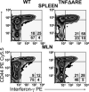
The proportion of CD8+/CD44high T cells that produce IFN-γ was increased in TNFΔARE mice. Lymphocytes isolated from indicated populations of TNFΔARE mice or WT littermates (WT) were cultured under PMA/ionomycin stimulation and incubated with Abs against CD8, CD44, and IFN-γ, as per manufacturer's instructions, and then were analyzed by flow cytometry. Cells were gated on FSC, SSC, and CD8 using CD44-deficient lymphocytes for surface staining and isotype Ab for intracellular staining (MFI < 101; data not shown). Representative density plots were obtained from three experiments using three to four mice per strain at ≥20 wk of age and run in duplicate.
FIGURE 5
FIGURE 5
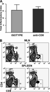
Depletion of CD8+ T cells did not affect the severity of ileitis in TNFΔARE mice. A, Eight-week-old TNFΔARE mice received five injections of a CD8-depleting Ab or a corresponding isotype Ab control every 2 days. Ileal tissues were harvested 2 days after the last injection, and the severity of ileitis was assessed as described (mean ± SEM, n = 7/group). B, Depletion of CD8+ T cells was confirmed by flow cytometry from peripheral blood at treatment day 6 and from MLN and spleen at day 12 (representative plots).
FIGURE 6
FIGURE 6
CD8+ T cells polarize into CD44high/CD103low and CD44low/CD103high populations in TNFΔARE mice. Lymphocytes isolated from indicated organs of TNFΔARE mice or WT littermates were incubated with Abs against CD8, CD44, CD103, and L-selectin and analyzed by flow cytometry. Cells were gated on FSC, SSC, and CD8+, and right panels were additionally gated on CD103. Representative density plots were obtained from three to four mice per strain at ≥20 wk of age run in duplicate or triplicate.
FIGURE 7
FIGURE 7
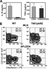
CD8+/CD103high T cells from TNFΔARE mice produce TGF-β, but not IL-10 or IFN-γ. A, CD4+ T cells were isolated from the MLN and spleen of TNFΔARE mice using magnetic beads and used as positive controls for cytokine production. The CD4negative fraction was then sorted by FACS as per the strategy illustrated in Fig. 8 (top density plots). The CD8+/CD103high and CD4+ fractions were cultured as described, and cytokine concentrations in the supernatants of cultured cells (mean ± SEM from two experiments) pooled from six mice per experiment, run in triplicate, as per Materials and Methods. B, Lymphocytes isolated from the indicated populations of TNFΔARE mice or WT littermates were incubated with Abs against CD8, CD103, and IFN-γ and analyzed by flow cytometry. Cells were gated on FSC, SSC, and CD8 using CD103-deficient lymphocytes for surface staining and isotype Ab for intracellular staining (MFI < 101; data not shown). Representative density plots were obtained from three experiments using three to four mice per strain at ≥20 wk of age and run in duplicate.
FIGURE 8
FIGURE 8
CD8+/CD103high, but not CD8+/CD44 high T cells isolated from TNFΔARE mice decreased proliferation of CD4+ T cells more efficiently than CD8+/CD103high T cells isolated from WT littermates. A and B, CD4+ T cells from TNFΔARE mice were isolated magnetically, sorted by flow cytometry as delineated in the top plots, and cocultured with APCs in the presence or absence of CD8+/CD103high or CD8+/CD44high T cells from TNFΔARE or WT mice. Incorporation of [3H]thymidine was determined, as described in Materials and Methods. Data are presented as mean ± SEM from two independent experiments run in trip-licate; *, p < 0.001.
FIGURE 9
FIGURE 9
CD8+/CD103high T cells isolated from WT or TNFΔARE mice reconstituted the CD8+ compartment of RAG–/– mice and attenuated the ileitis induced by adoptive transfer of CD4+ T cells from TNFΔARE mice. A–D, CD4+ T cells were transferred alone (A and D)or with CD8+/CD103high (B–D) or CD8+/CD44high (D) T cells (sorted as illustrated in Fig. 8) into RAG–/– mice. The reconstitution of the subsets and the effect on the severity of ileitis were assessed after 6 wk, as previously described (mean ± SEM from two experiments, n = 7–14/group; *, p < 0.05). E and F, Representative histology of the terminal ileum of mice transferred with indicated T cell subsets (representative micrographs; H&E; original magnification ×20).

References

    1. Izcue A, Coombes JL, Powrie F. Regulatory T cells suppress systemic and mucosal immune activation to control intestinal inflammation. Immunol. Rev. 2006;212:256–271. - PubMed
    1. Mottet C, Uhlig HH, Powrie F. Cutting edge: cure of colitis by CD4+CD25+ regulatory T cells. J. Immunol. 2003;170:3939–3943. - PubMed
    1. Powrie F, Read S, Mottet C, Uhlig H, Maloy K. Control of immune pathology by regulatory T cells. Novartis Found. Symp. 2003;252:92–98. - PubMed
    1. Strober W, Fuss IJ, Blumberg RS. The immunology of mucosal models of inflammation. Annu. Rev. Immunol. 2002;20:495–549. - PubMed
    1. Fiocchi C. Inflammatory bowel disease: etiology and pathogenesis. Gastroenterology. 1998;115:182–205. - PubMed

Publication types

MeSH terms