

Application of light microscopical and ultrastructural immunohistochemistry in the study of goblet cell carcinoid in the appendix
- PMID: 18252007
- PMCID: PMC2275273
- DOI: 10.1186/1477-7819-6-15
Application of light microscopical and ultrastructural immunohistochemistry in the study of goblet cell carcinoid in the appendix
Abstract
Background: Goblet cell carcinoids appear less frequently in the appendix than do other carcinoids. In the presented work a case with a goblet cell carcinoid of the appendix is described.
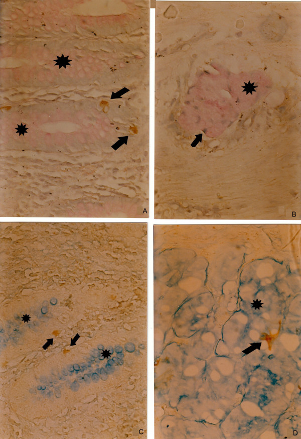
Methods: Routine histological and histochemical methods were employed, with a combination of histochemistry and immunohistochemistry on one section and light and electron microscopical immunohistochemisty on paraffin-embedded material, were applied to identify the type of the carcinoid and to reveal the fine structure of cell types in the tumour nests of the appendix.
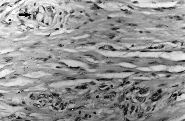
Results: During the biopsy of a patient who had undergone appendectomy, an infiltration with clusters of goblet cells in the submucosa of the appendix was found. After a second operation of right-sided hemicolectomy, similar clusters of goblet cells were detected in the muscle layers of the caecum. After 18 months the patient died from cirrhosis and had not developed metastases or any recurrence. Immunohistochemically the serotonin-, somatostatin-, chromogranin A- and synaptophysin-positive endocrine cells were basally attached to mucin-secreting cells. The combined staining revealed simultaneously present endocrine cells (chromogranin-A-positive) and mucin-secreting cells (PAS- or alcian blue-positive). The ultrastructural immunohistochemistry showed that chromogranin A-positive cells had discoid and pleomorphic granules and were located in tumour nests or as single cells in the appendiceal wall.
Conclusion: The combined histochemical and immunohistochemical procedure and the ultrastructural immunohistochemistry on archival material could contribute in clarifying the diagnosis of goblet cell carcinoid.
Figures


References
-
- Burke AP, Sobin LH, Federspiel BH, Shekitka KM, Helwig EB. Goblet cell carcinoids and related tumors of the vermiform appendix. Am J Clin Pathol. 1990;94:27–35. - PubMed
-
- Capella C, La Rosa S, Uccella S, Billo P, Cornaggia M. Mixed endocrine-exocrine tumors of the gastrointestinal tract. Semin Diagn Pathol. 2000;17:91–103. - PubMed
-
- Oberg K, Astrup L, Eriksson B, Falkmer SE, Falkmer UG, Gustafsen J, Haglund C, Knigge U, Vatn MH, Valimaki M. Guidelines for the management of gastroenteropancreatic neuroendocrine tumours (including bronchopulmonary and thymic neoplasms). Part I-general overview. Acta Oncol. 2004;43:617–625. doi: 10.1080/02841860410018575. - DOI - PubMed
-
- Burke AP, Sobin LH, Federspiel BH, Shekitka KM. Appendiceal carcinoids: correlation of histology and immunohistochemistry. Mod Pathol. 1989;2:630–637. - PubMed
Publication types
MeSH terms
Substances
LinkOut - more resources
Full Text Sources
Medical
Research Materials
