. 2008 Feb;82(2):432-43.
doi: 10.1016/j.ajhg.2007.11.002. Epub 2008 Jan 24.

Submicroscopic duplications of the hydroxysteroid dehydrogenase HSD17B10 and the E3 ubiquitin ligase HUWE1 are associated with mental retardation

Affiliations

Submicroscopic duplications of the hydroxysteroid dehydrogenase HSD17B10 and the E3 ubiquitin ligase HUWE1 are associated with mental retardation

Guy Froyen et al. Am J Hum Genet. 2008 Feb.

Abstract

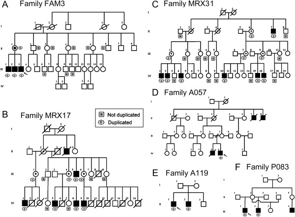
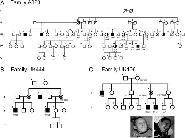
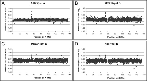
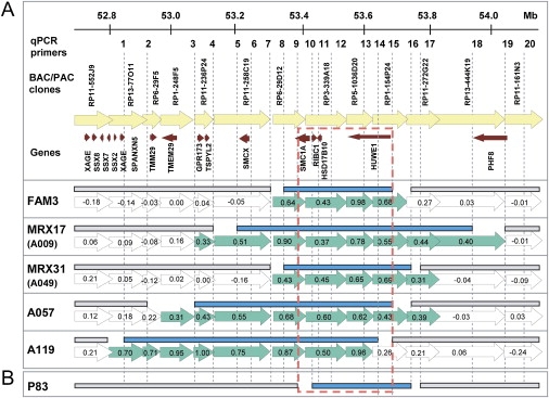
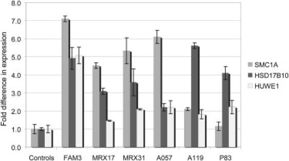
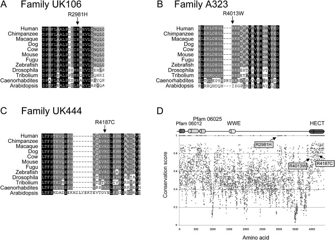
Submicroscopic copy-number imbalances contribute significantly to the genetic etiology of human disease. Here, we report a novel microduplication hot spot at Xp11.22 identified in six unrelated families with predominantly nonsyndromic XLMR. All duplications segregate with the disease, including the large families MRX17 and MRX31. The minimal, commonly duplicated region contains three genes: RIBC1, HSD17B10, and HUWE1. RIBC1 could be excluded on the basis of its absence of expression in the brain and because it escapes X inactivation in females. For the other genes, expression array and quantitative PCR analysis in patient cell lines compared to controls showed a significant upregulation of HSD17B10 and HUWE1 as well as several important genes in their molecular pathways. Loss-of-function mutations of HSD17B10 have previously been associated with progressive neurological disease and XLMR. The E3 ubiquitin ligase HUWE1 has been implicated in TP53-associated regulation of the neuronal cell cycle. Here, we also report segregating sequence changes of highly conserved residues in HUWE1 in three XLMR families; these changes are possibly associated with the phenotype. Our findings demonstrate that an increased gene dosage of HSD17B10, HUWE1, or both contribute to the etiology of XLMR and suggest that point mutations in HUWE1 are associated with this disease too.


Figures

Figure 1
Figure 1
Pictures of the Three Affected Individuals of FAM3 with a Duplication at Xp11.22 Facial features of the sons (from left to right; III-3, III-2, and III-1). Note hypertelorism, short and upslanting palpebral fissures, epicanthi, lateral flare of eyebrows, broad nose, simple protruding ears, open mouth, and thick lips.
Figure 2
Figure 2
Pedigrees of Families with a Microduplication at Xp11.22 (A) FAM3, (B) MRX17, (C) MRX31, (D) A057, (E) A119, and (F) P083. Family members that could be tested for the duplication are indicated as N (do not carry the duplication) or D (carry the duplication).
Figure 3
Figure 3
Pedigrees of Families with a Sequence Change in HUWE1 (A) A323, (B) UK444, and (C) UK106. Additional family members that were screened are indicated on the pedigrees as WT or WT/WT (do not carry the likely mutation), mut (carry the mutation), or WT/mut (heterozygous for the mutation). For the large family A323, the HUWE1 gene is located within the linked interval. Female I.2 of family UK106 most probably is germline mosaic. Pictures of the affected individual III-6 and III-7 of family UK106 are shown.
Figure 4
Figure 4
X Chromosome-Specific Array-CGH Plots Obtained for Probands of Four Families Revealing the Duplications at Xp11.22 These families are (A) FAM3, (B) MRX17, (C) MRX31, and (D) A057. DNA from two unrelated MR patients were differentially labeled and cohybridized onto the X array. The log2 normalized intensity ratios of the Cy5 (patient with duplication) and Cy3 (unrelated MR patient, named pat A–D) signals is plotted (y axis) against the position on the X chromosome (in Mb), from Xpter to Xqter (x axis). The duplications are visible as clones with aberrant ratios >0.3 at ∼53 Mb. For A057, an additional polymorphic duplication is visible as three aberrant clones at 38 Mb. Other clones outside the normal interval (log2 ratio of −0.3 to 0.3) are polymorphic clones or outliers.
Figure 5
Figure 5
Schematic Representation of the Xp11.22 Region Showing the Duplications (A) Indicated from top to bottom: the location of the qPCR primer sets (1–20), genomic BAC or PAC clones, and annotated genes present in the interval composed of the six duplications. This region is represented for each family by the arrows indicating the genomic clones from the X array with their corresponding log2 ratios. Clones with aberrant ratios are in green; those with ratios in the normal interval are open. For each family, the mapped duplicated region, defined by qPCR, is given as solid blue bars above these clones, indicating that the primer sets within these bars yielded relative copy numbers of ∼2.0 whereas the primer pairs outside (indicated with the gray bars) gave values ∼1.0. Proximal and distal breakpoints must lie between these bars (no bars). (B) For P083, only qPCR data are shown. The maximal common duplicated region is boxed (broken red line) and entirely contains the genes RIBC1, HSD17B10, and HUWE1.
Figure 6
Figure 6
Representative FISH Data on Chromosomes from Probands of Families MRX17, MRX31, and A057 (A) Labeled DNA of RP6-29D12 (red) and RP1-154P24 (green) were hybridized to metaphase spreads and show signals for both probes only at Xp11. (B) On interphase chromosomes, alternating red-green-red-green signals are indicative of a tandem duplication event.
Figure 7
Figure 7
mRNA Expression Analysis of SMC1A, HSD17B10, and HUWE1 in All Six Families with a Duplication at Xp11.22 cDNA was prepared from RNA extracted from blood (controls and FAM3) or EBV-transformed PBL cell lines (controls, MRX17, MRX31, A057, A119, and P083). Compared to controls, all affected individuals showed significantly increased mRNA levels for all three genes except for SMC1A in family P083. Expression was determined by real-time RT-PCR with the comparative ddCt method and normalized to the expression of HPRT and ACTB (FAM3, P083) or with the standard curve method with ACTB for normalization (MRX17, MRX31, A057, and A119). Expression levels are calculated relative to the mean levels obtained in the control samples (fold difference; y axis). Standard deviations of at least two independent experiments are indicated for each bar.
Figure 8
Figure 8
Missense Changes in the HUWE1 Gene Identified in Three XLMR Families Partial ClustalW alignments of HUWE1 orthologs in the regions surrounding (A) R2981H (UK106), (B) R4013W (A323), and (C) R4187C (UK444) amino acid changes. Whereas the R2981 is invariable across all orthologs, R4013 and R4187 are highly conserved. In (D), the position of these arginine residues within the HUWE1 protein is indicated with the arrows. The cartoon above the conservation plot (Scorecons) indicates domains within the HUWE1 protein. Pfam 06012 (aa 104–374) and Pfam 06025 (aa 424–704 and 762–815) domains are of unknown function, WWE domain (aa 1617–1678) might be involved in the regulation of ubiquitin-mediated proteolysis, and HECT domain (aa 4036–4374) is the catalytic domain of HUWE1 ubiquitin protein ligase.

References

    1. Leonard H., Wen X. The epidemiology of mental retardation: Challenges and opportunities in the new millennium. Ment. Retard. Dev. Disabil. Res. Rev. 2002;8:117–134. - PubMed
    1. Gecz J. The molecular basis of intellectual disability: Novel genes with naturally occurring mutations causing altered gene expression in the brain. Front. Biosci. 2004;9:1–7. - PubMed
    1. Ropers H.H. X-linked mental retardation: Many genes for a complex disorder. Curr. Opin. Genet. Dev. 2006;16:260–269. - PubMed
    1. Albertson D.G., Pinkel D. Genomic microarrays in human genetic disease and cancer. Hum. Mol. Genet. 2003;12:R145–R152. - PubMed
    1. Vissers L.E., de Vries B.B., Osoegawa K., Janssen I.M., Feuth T., Choy C.O., Straatman H., van der Vliet W., Huys E.H., Van Rijk A. Array-based comparative genomic hybridization for the genome-wide detection of submicroscopic chromosomal abnormalities. Am. J. Hum. Genet. 2003;73:1261–1270. - PMC - PubMed

Publication types