. 2008 Feb;82(2):501-9.
doi: 10.1016/j.ajhg.2007.10.004. Epub 2008 Jan 31.

TINF2, a component of the shelterin telomere protection complex, is mutated in dyskeratosis congenita

Affiliations

TINF2, a component of the shelterin telomere protection complex, is mutated in dyskeratosis congenita

Sharon A Savage et al. Am J Hum Genet. 2008 Feb.

Abstract

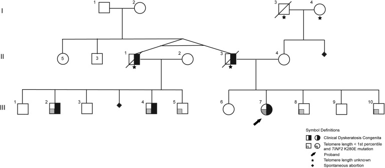
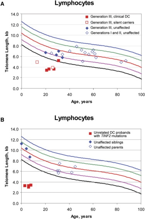
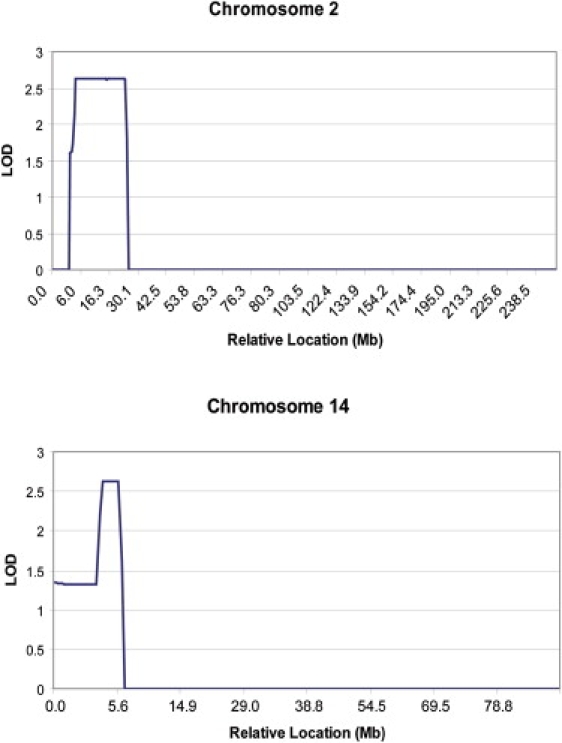
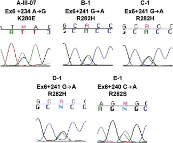
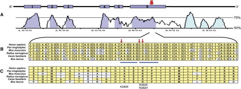
Patients with dyskeratosis congenita (DC), a heterogeneous inherited bone marrow failure syndrome, have abnormalities in telomere biology, including very short telomeres and germline mutations in DKC1, TERC, TERT, or NOP10, but approximately 60% of DC patients lack an identifiable mutation. With the very short telomere phenotype and a highly penetrant, rare disease model, a linkage scan was performed on a family with autosomal-dominant DC and no mutations in DKCI, TERC, or TERT. Evidence favoring linkage was found at 2p24 and 14q11.2, and this led to the identification of TINF2 (14q11.2) mutations, K280E, in the proband and her five affected relatives and TINF2 R282H in three additional unrelated DC probands, including one with Revesz syndrome; a fifth DC proband had a R282S mutation. TINF2 mutations were not present in unaffected relatives, DC probands with mutations in DKC1, TERC, or TERT or 298 control subjects. We demonstrate that a fifth gene, TINF2, is mutated in classical DC and, for the first time, in Revesz syndrome. This represents the first shelterin complex mutation linked to human disease and confirms the role of very short telomeres as a diagnostic test for DC.


Figures

Figure 1
Figure 1
Pedigree of Family A The monozygotic twins in generation II had five sisters and three brothers. Telomere length was determined on individuals except where indicated by a star.
Figure 2
Figure 2
Lymphocyte Telomere Lengths (A) Lymphocyte telomere lengths measured by flow-FISH of family A, including generation III affected individuals (red filled squares), generation III silent carriers (red open squares), unaffected members of generation III (blue filled diamonds), and unaffected members of generations I and II (blue open diamonds). The black line indicates the first percentile, and the blue line indicates the 99th percentile. Individual III-05 (filled red square on the first percentile line) had telomere lengths of 5.2 kb. The first percentile for age is 5 kb. However, his telomere lengths were less than the first percentile for age in naive T cells and granulocytes (data not shown). (B) Lymphocyte telomere lengths of the four unrelated DC probands (filled red circles) with TINF2 mutations, their unaffected parents (open blue diamonds), and unaffected siblings (filled blue diamonds).
Figure 3
Figure 3
LOD Scores for Chromosomes 2 and 14 LOD scores were determined under an autosomal-dominant (parametric), rare disease model with telomere length less than the first percentile as the affected phenotype as described in the text. The relative location of the markers is noted on the x axis. (Top) A total of 356 SNP markers on chromosome 2 were evaluated after removal of SNPs in linkage disequilibrium as described. The maximum LOD score of 2.62 was located between SNP markers rs6767 and rs520354 on chromosome 2 at positions 3.5 Mb and 21.1 Mb, respectively. DDX1 was the best candidate gene in this location. (Bottom) A total of 161 SNP markers on chromosome 14 were evaluated after removal of SNPs in linkage disequilibrium as described. The maximum LOD score of 2.62 was located between SNP markers rs1570342 and rs195677 on chromosome 14 at positions 22.4 Mb and 25.2 Mb, respectively. TINF2 was the best candidate gene in this location.
Figure 4
Figure 4
Representative Sequence Tracings of TINF2 Mutations The proband and mutation are noted above the sequence tracing.
Figure 5
Figure 5
Genomic Structure and Evolutionary Conservation of TINF2 (A) The genomic region of TINF2 consists of 2686 base pairs. The red arrows show the relative positions of the mutations identified in the DC patients in this study, Ex6+234A→G (K280E), Ex6+240C→A (R282S), and Ex6+241G→A (R282H). Curves showing conservation between human and mouse genomic sequences were generated with VISTA. On the right, 50% and 75% conservation are noted. Exons are shown in blue, and the conserved 3′ untranslated region is shown in light blue. (B) Comparison of nucleotide conservation between Homo sapiens (Entrez Gene Annotation, NC_000014.7), Pan troglodytes (NC_006481.2), Mus musculus (NC_00080.5), Rattus norvegicus (NC_005114.2), Canis lupus familiaris (NC_006590.2), and Bos taurus (NC_007308.2) TINF2 genomic sequences between Ex6+204 through Ex6+272. Conserved nucleotides are shown in yellow. Mutated nucleotides are noted with red arrows. The blue lines above the nucleotides indicate putative exonic splice enhancer (ESE) sequences. (C) Comparison of amino acid conservation between Homo sapiens (Entrez Protein Annotation, NP_036593.1), Pan troglodytes (CAH89509.1, hypothetical protein TIN2), Mus musculus (NP_663751.2), Rattus norvegicus (NP_001006963.1), Canis lupus familiaris (XP_850486.1, predicted protein), and Bos taurus (XP_593926.3, predicted protein) between amino acids 270 and 292. Conserved amino acids are noted in yellow. Mutated amino acids K280E, R282S, and R292H are noted at the bottom.

References

    1. Walne A.J., Marrone A., Dokal I. Dyskeratosis congenita: A disorder of defective telomere maintenance? Int. J. Hematol. 2005;82:184–189. - PubMed
    1. Vulliamy T.J., Marrone A., Knight S.W., Walne A., Mason P.J., Dokal I. Mutations in dyskeratosis congenita: Their impact on telomere length and the diversity of clinical presentation. Blood. 2006;107:2680–2685. - PubMed
    1. Tamary H., Alter B.P. Current diagnosis of inherited bone marrow failure syndromes. Pediatr. Hematol. Oncol. 2007;24:87–99. - PubMed
    1. Sznajer Y., Baumann C., David A., Journel H., Lacombe D., Perel Y., Blouin P., Segura J.F., Cezard J.P., Peuchmaur M. Further delineation of the congenital form of X-linked dyskeratosis congenita (Hoyeraal-Hreidarsson syndrome) Eur. J. Pediatr. 2003;162:863–867. - PubMed
    1. Revesz T., Fletcher S., al Gazali L.I., DeBuse P. Bilateral retinopathy, aplastic anaemia, and central nervous system abnormalities: A new syndrome? J. Med. Genet. 1992;29:673–675. - PMC - PubMed

Publication types

Substances