. 2008 May 10;374(2):280-91.
doi: 10.1016/j.virol.2007.12.039. Epub 2008 Feb 5.

Protein-RNA tethering: the role of poly(C) binding protein 2 in poliovirus RNA replication

Affiliations

Protein-RNA tethering: the role of poly(C) binding protein 2 in poliovirus RNA replication

Allyn Spear et al. Virology. .

Abstract

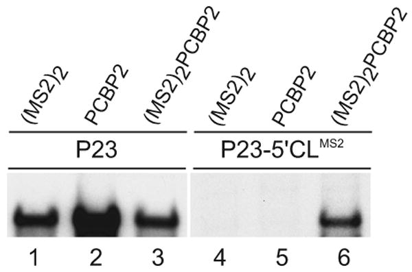
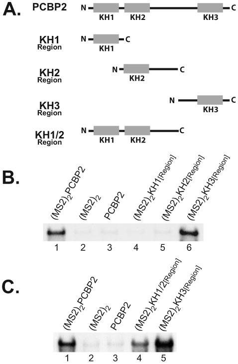
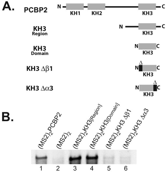
The exploitation of cellular functions and host proteins is an essential part of viral replication. The study of this interplay has provided significant insight into host cell processes in addition to advancing the understanding of the viral life-cycle. Poliovirus utilizes a multifunctional cellular protein, poly(C) binding protein 2 (PCBP2), for RNA stability, translation and RNA replication. In its cellular capacity, PCBP2 is involved in many functions, including transcriptional activation, mRNA stability and translational silencing. Using a novel protein-RNA tethering system, we establish PCBP2 as an essential co-factor in the initiation of poliovirus negative-strand synthesis. Furthermore, we identified the conserved KH domains in PCBP2 that are required for the initiation of poliovirus negative-strand synthesis, and showed that this required neither direct RNA binding or dimerization of PCBP2. This study demonstrates the novel application of a protein-RNA tethering system for the molecular characterization of cellular protein involvement in viral RNA replication.


Figures

Fig. 1
Fig. 1
Schematic of poliovirus RNAs used in this study. (A) PV1ΔGUA3 RNA contains the entire poliovirus genomic RNA sequence with a five nucleotide deletion in the 3′NTR, rendering it incapable of RNA replication. P23 RNA contains a deletion of the P1 capsid coding region, however, it does encode all essential viral replication proteins. F3 RNA contains a deletion of both the P1 and P2 coding regions. In addition, F3 RNA contains a two nucleotide deletion at the beginning of the P3 coding region, producing a frameshift which results in premature termination of translation. (B) Sequence diagrams of both the wild-type 5′ cloverleaf with the C24A mutation (left) and the MS2 5′ cloverleaf (right).
Fig. 2
Fig. 2
A mutation affecting PCBP2 binding to the 5′ cloverleaf affects negative-strand synthesis. Replication of subgenomic RNAs were measured using preinitiation replication complexes (PIRCs) isolated from HeLa S10 translation-replication reactions. Radiolabeled product RNAs were visualized by CH3HgOH-agarose gel electrophoresis and autoradiography. (A) Replication of wild-type and 5′CL(C24A) RNAs were assayed as described in Materials and Methods. P23 RNA transcripts contained two additional 5′ terminal G residues and support only negative-strand synthesis. RzP23 RNA has an authentic 5′ terminus that was generated by the inclusion of a hammerhead ribozyme upstream of the first viral nucleotide, and supports both negative- and positive-strand synthesis. (B) Replication of either wild-type F3 RNA or F3-5′CL(C24A) RNA was performed in trans-replication assays as described in Materials and Methods. A non-replicating helper RNA [PV1ΔGUA3] was used to provide equal levels of the poliovirus replication proteins in each reaction. Each input template RNA was capped, to ensure equal template stability.
Fig. 3
Fig. 3
Schematic of (MS2)2 protein tethering system. (A) Template RNAs which contain the wild-type 5′CL bind PCBP2 at stem-loop b and viral protein 3CD at stem-loop d. (B) Template RNAs which contain the mutant 5′CLMS2 (Fig. 1B) do not bind PCBP2, but do bind viral protein 3CD. (MS2)2PCBP2 fusion protein binds to the MS2 stem-loop in the 5′CLMS2 RNA via the (MS2)2-RNA interaction.
Fig. 4
Fig. 4
(MS2)2PCBP2 binds selectively to the mutant 5′CLMS2 RNA. (A) Electrophoretic mobility shift assay using radiolabeled RNA probes, either 5′CLWT RNA (lanes 1-4) or 5′CLMS2 RNA (lanes 5-8). The RNA probe was either run alone (lanes 1 & 5), with mock HeLa S10 translation reaction (lanes 2 & 6), with bacterially expressed rPCBP2 (lanes 3 & 7) or a vector control bacterial extract (lanes 4 & 8). The specific RNP complex formed with the 5′CLWT RNA probe and endogenous cellular PCBP is labeled as complex I. (B) Electrophoretic mobility shift assay using radiolabeled RNA probes, either 5′CLWT RNA (lanes 1-5) or 5′CLMS2 RNA (lanes 6-10). The probe was either run alone (lanes 1 & 6), with a mock HeLa S10 translation reaction (lanes 2 & 7), or with HeLa S10 translation reactions in which the indicated proteins were expressed (lanes 3-5, 8-10). Specific RNP complexes were formed with the 5′CLWT RNA and endogenous cellular PCBP (complex I), or with the 5′CLMS2 RNA and (MS2)2 or (MS2)2PCBP2 (complex II).
Fig. 5
Fig. 5
Expression of (MS2)2PCBP2 restores negative-strand synthesis on a 5′CLMS2 RNA template. Negative-strand synthesis was measured in reactions containing either P23 RNA (lanes 1-3) or P23-5′CLMS2 RNA (lanes 4-6) as described in Materials and Methods. Each reaction contained either P23 RNA or P23-5′CLMS2 RNA and an equimolar amount of a protein expression RNA which expressed either (MS2)2, PCBP2 or (MS2)2PCBP2, as indicated. Both template RNAs were capped to ensure equal template stability.
Fig. 6
Fig. 6
Identification of the KH domains in PCBP2 that are required for negative-strand synthesis using the (MS2)2 protein-RNA tethering system. (A) Schematic of the domain structure of PCBP2 (top), including the conserved KH1, KH2 and KH3 domains. PCBP2 was divided into three regions based on the KH domains, and the resulting protein fragments are depicted above. An N-terminal fragment of PCBP2 which contained both the KH1 and KH2 domains is also shown. (B) Negative-strand synthesis was measured in reactions containing P23-5′CLMS2 RNA, and an equimolar amount of a protein expression RNA which expressed either (MS2)2, PCBP2, (MS2)2PCBP2, (MS2)2 KH1[Region], (MS2)2 KH2[Region], (MS2)2 KH3[Region] or (MS2)2 KH1/2[Region] as indicated. (C) Negative-strand synthesis was measured in reactions containing P23-5′CLMS2 RNA and a protein expression RNA as indicated above. The total molar RNA concentration and molar RNA ratio were maintained in each reaction, and the input template RNA contained a 5′ cap.
Fig. 7
Fig. 7
Levels of protein synthesis observed in the (MS2)2 protein-RNA tethering replication reactions. HeLa S10 translation reactions which correlate to those described in Fig. 6 were incubated with [35S]methionine label the protein products. The labeled proteins synthesized in these reactions were analyzed by SDS-PAGE and autoradiography. Each reaction contained an equimolar amount of P23-5′CLMS2 RNA and the indicated (MS2)2 fusion protein expression RNA.
Fig. 8
Fig. 8
Characterization of the PCBP2 KH3 domain in negative-strand synthesis using the (MS2)2 protein-RNA tethering system. (A) Schematic of PCBP2 and the KH3 domain deletion mutants used in this experiment. (B) Negative-strand synthesis was measured in reactions containing P23-5′CLMS2 RNA and an equimolar amount of a protein expression RNA as indicated above. The total molar RNA concentration and molar RNA ratio were maintained in each reaction, and the input template RNA contained a 5′ cap.
Fig. 9
Fig. 9
The (MS2)2 fusion proteins bind to 5′CLMS2 RNA with equal affinity. (A) Electrophoretic mobility shift assays were performed using a radiolabeled 5′CLMS2 RNA probe. The probe was either run alone (lane 1), with a mock HeLa S10 translation reaction (lane 2), or with HeLa S10 translation reactions in which the indicated proteins were expressed (lanes 3-11). (B) The same HeLa S10 translation reactions used above were also incubated with [35S]methionine, and the labeled protein products were analyzed by SDS-PAGE and autoradiography.
Fig. 10
Fig. 10
Model of circular RNA replication complex formation with PCBP2 and (MS2)2PCBP2 in cell-free reactions. (A) Current model showing formation of circular RNP complex required to initiate negative-strand synthesis. Cellular proteins PCBP and PABP bind to the 5′ and 3′ ends of PV RNA respectively, forming a protein-protein bridge which circularizes PV genomic RNA. Proteolytic cleavage of 3CD at the 3′ end generates active 3D polymerase which utilizes viral protein VPg to initiate negative-strand RNA synthesis. (B) Modified model in which (MS2)2PCBP2 tethered to 5′CLMS2 RNA in place of PCBP2 is capable of forming the same requisite interactions for genome circularization and the initiation of negative-strand synthesis.

Similar articles

Cited by

References

    1. Andino R, Rieckhof GE, Achacoso PL, Baltimore D. Poliovirus RNA synthesis utilizes an RNP complex formed around the 5′-end of viral RNA. EMBO J. 1993;12:3587–3598. - PMC - PubMed
    1. Andino R, Rieckhof GE, Baltimore D. A functional ribonucleoprotein complex forms around the 5′ end of poliovirus RNA. Cell. 1990;63:369–380. - PubMed
    1. Barton DJ, Black EP, Flanegan JB. Complete replication of poliovirus in vitro: Preinitiation RNA replication complexes require soluble cellular factors for the synthesis of VPg-linked RNA. The Journal of Virology. 1995;69:5516–5527. - PMC - PubMed
    1. Barton DJ, Morasco BJ, Flanegan JB. Assays for poliovirus polymerase, 3Dpol, and authentic RNA replication in HeLa S10 extracts. Methods Enzymol. 1996;275:35–57. - PubMed
    1. Barton DJ, Morasco BJ, Flanegan JB. Translating ribosomes inhibit poliovirus negative-strand RNA synthesis. The Journal of Virology. 1999;73:10104–10112. - PMC - PubMed

Publication types