. 2008 Apr 18;283(16):10978-91.
doi: 10.1074/jbc.M800622200. Epub 2008 Feb 5.

Glycine transporter dimers: evidence for occurrence in the plasma membrane

Affiliations

Glycine transporter dimers: evidence for occurrence in the plasma membrane

Ingo Bartholomäus et al. J Biol Chem. .

Abstract

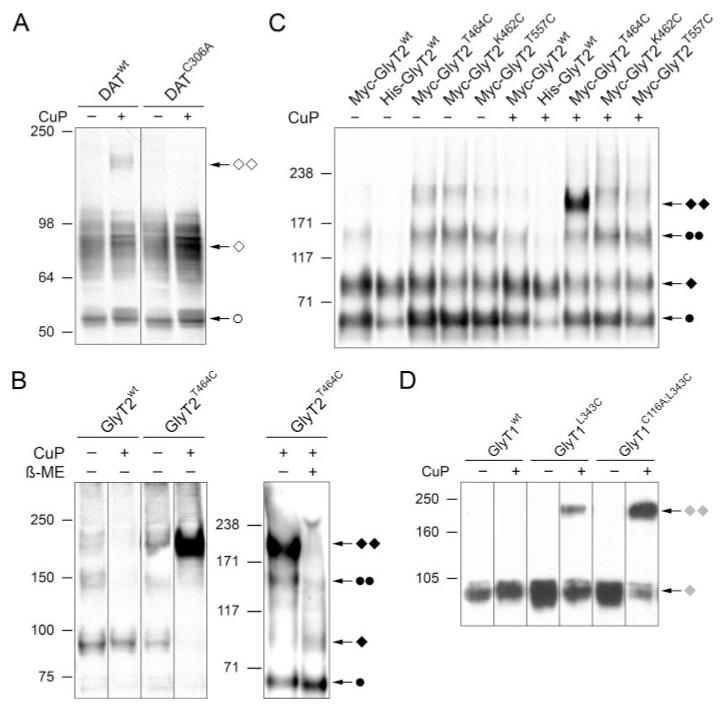
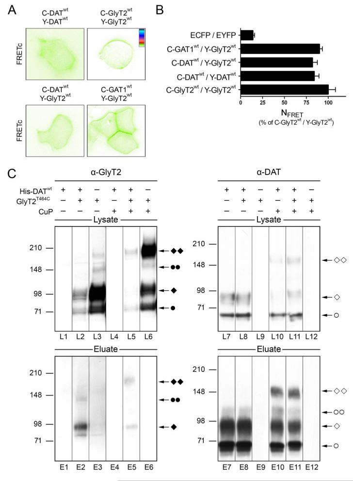
Different Na(+)/Cl(-)-dependent neurotransmitter transporters of the SLC6a family have been shown to form dimers or oligomers in both intracellular compartments and at the cell surface. In contrast, the glycine transporters (GlyTs) GlyT1 and -2 have been reported to exist as monomers in the plasma membrane based on hydrodynamic and native gel electrophoretic studies. Here, we used cysteine substitution and oxidative cross-linking to show that of GlyT1 and GlyT2 also form dimeric complexes within the plasma membrane. GlyT oligomerization at the cell surface was confirmed for both GlyT1 and GlyT2 by fluorescence resonance energy transfer microscopy. Endoglycosidase treatment and surface biotinylation further revealed that complex-glycosylated GlyTs form dimers located at the cell surface. Furthermore, substitution of tryptophan 469 of GlyT2 by an arginine generated a transporter deficient in dimerization that was retained intracellulary. Based on these results and GlyT structures modeled by using the crystal structure of the bacterial homolog LeuT(Aa), as a template, residues located within the extracellular loop 3 and at the beginning of transmembrane domain 6 are proposed to contribute to the dimerization interface of GlyTs.


Figures

FIGURE 1
FIGURE 1. Model of the structure of GlyT2
A, alignment of partial amino acid sequences of GlyT1, GlyT2, DAT, and LeuTAa. B–E, homology model of GlyT2 based on the crystal structure of LeuTAa. B and C, the putative GlyT2 dimer is depicted from the extracellular side (B) and within the plane of the membrane (C). Helices (cylinders) are numbered following the LeuTAa nomenclature (13). D and E, close-up views of the dimer interface shown in B and C. Residues substituted in this study are indicated.
FIGURE 2
FIGURE 2. Oxidative cross-linking of cysteine-substituted GlyTs
A, Western blot of detergent extracts of HEK 293T cells expressing DATWT or DATC306A. The cells were treated with PBS (−) or CuP (+), prior to SDS-PAGE and immunoblotting with a DAT-specific antibody. Bands assigned to the immature monomer (○), the mature monomer (◇), and its dimer (◇◇) are indicated. B (left), HEK 293T cells were transfected with GlyT2WT or GlyT2T464C and treated with CuP (+), or PBS (−), prior to detergent extraction and Western blot analysis with a GlyT2-specific antibody. Right, detergent extracts from CuP-treated cells expressing GlyT2T464C were supple mented withβ-mercaptoethanol (100 mm) or left untreated, prior to Western blot analysis with a GlyT2-specific antibody. C, Western blot of detergent extracts prepared from HEK 293T cells expressing Myc- or His-tagged GlyT2WT and the Myc-GlyT2T464C, Myc-GlyTK462C, or Myc-GlyT2T557C mutants. Prior to detergent extraction, cells were treated with CuP (+) or PBS (−), as indicated. D, GripTite 293 cells expressing FLAG-tagged GlyT1, GlyT1L343C, or GlyT1C116A/L343C were treated with CuP (+) or PBS (−), prior to detergent extraction and Western blot analysis with an anti-FLAG antibody. Note that both GlyTs formed adducts of higher molecular weight upon treatment with CuP only when cysteine-substituted. Different transporter forms are indicated: ●, immature monomer; ◆, mature monomer; ●●, immature dimer; ◆ ◆, mature dimer. Filled symbols, GlyT2; shaded symbols, GlyT1.
FIGURE 3
FIGURE 3. The CuP-induced GlyT2 adduct is complex-glycosylated and localized at the cell surface
A, detergent extracts of CuP (+)-treated HEK 293T cells transfected with His-GlyT2WT or His-GlyT2T464C were incubated with endoglycosidase H (Endo H) or peptide:N-glycosidase F (PNGase F), as indicated, and analyzed by Western blotting using a GlyT2-specific antibody. B, HEK 293T cells transfected with His-GlyT2WT or His-GlyT2T464C were treated with CuP (+), washed, and subjected to surface biotinylation. Aliquots of the detergent extracts prepared from these cells (total) as well as the eluates from the streptavidin-agarose beads containing the biotinylated proteins (surface) were analyzed by Western blotting analysis with GlyT2-specific antibodies. Different transporter forms are indicated as follows. ●, immature monomer; ⊗, deglycosylated monomer; ◆, mature monomer; ●●, immature dimer; ⊗⊗, deglycosylated dimer; ◆ ◆, mature dimer.
FIGURE 4
FIGURE 4. GlyT oligomerization demonstrated by FRET in intact cells
A, HEK 293T cells were transiently transfected with cDNAs encoding CFP- or YFP-tagged proteins as indicated. Two days after transfection, epifluorescence microscopy was performed; the first and second columns show images obtained with CFP and YFP filter sets, and the third column displays a corrected and inverted FRET image (FRETc). A look-up table of the color code used is presented in the first and fourth image, third column (negative and positive control, respectively). All images shown are representative of 3–7 experiments. In all images, background fluorescence was subtracted. Scale bar, 10 μm. B, corrected and inverted FRET images of HEK 293T cells transiently transfected with the indicated constructs. For a look-up table of the color code used, see Fig. 5A. Images are representatives of at least three experiments. C, normalized FRET efficiencies (NFRET values) are given for cells expressing the following constructs: enhanced cyan fluorescent protein (ECFP) and enhanced yellow fluorescent protein (EYFP) (n = 124), C-GlyT1 and D2R-Y (n = 79), C-GlyT2 and D2R-Y (n = 50), C-GlyT2b and membrane-bound YFP (membr. YFP; n = 54), C-GlyT1 and Y-GlyT1 (n = 102), and C-GlyT2 and Y-GlyT2 (n = 73), respectively. Similarly, HEK 293 cells expressing C-DAT and Y-DAT (n = 30) as well as C-SERT-Y (n = 102; see also Ref. 37) were examined. Data represent means ± S.E.; ***, p < 0.001; analysis of variance, post hoc Bonferroni’s test for multiple comparisons.
FIGURE 5
FIGURE 5. The 200-kDa CuP-induced GlyT2 adduct is a homodimer
HEK 293T cells cotransfected with His-GlyT2T464C and either Myc-GlyT2WT or Myc-GlyT2T464C were treated with CuP (+) or PBS (−) as indicated. A, aliquots of detergent extracts prepared from the treated cells were sequentially analyzed by Western blotting using the GlyT2-specific antibody (lanes C1, C2, and L1–L8) and, after removal of bound antibody, an anti-Myc antibody (lanes L9–L16). Note the presence of mature GlyT2 dimers in lanes L6–L8 and L14 and L16, respectively. B, aliquots of the extracts used in A were affinity-purified on Ni2+-NTA-agarose. Blots prepared from the eluates were first probed with the GlyT2 antibody (lanes E1–E8) and, after removal of the bound antibodies, restained with a Myc-specific antibody (lanes E9–E16). Note that a Myc-immunoreactive 200-kDa adduct was isolated only after CuP treatment of cells expressing both His-GlyT2T464C and Myc-GlyT2T464C (lane E16). The different transporter forms are indicated: ●, immature monomer; ◆, mature monomer; ●●, immature dimer; ◆ ◆, mature dimer. An asterisk indicates an unspecific band detected with the anti-Myc antibody.
FIGURE 6
FIGURE 6. Heteromer formation of GlyT2 with DAT and GAT1
A, HEK 293T cells were transiently transfected with cDNAs encoding CFP- or YFP-tagged proteins, as indicated. Two days after transfection, FRETc values were obtained. A look-up table of the color code used is given in the upper right. NFRET values (percentage) normalized to C-GlyT2/Y-GlyT2-expressing cells (100%) are given for cells expressing ECFP and EYFP (n = 16), C-GATWT and Y-GlyT2 (n = 34), C-DATWT and Y-GlyT2WT (n = 34), C-DATWT and Y-DATWT (n = 30), and C-GlyT2WT and Y-GlyT2WT (n = 18), respectively. Note that NFRET values obtained from C-GlyT2WT/Y-GlyT2WT-expressing cells were not significantly different statistically from that of cells expressing C-GATWT/YGlyT2WT or C-DATWT/Y-GlyT2WT (p > 0.05). Data represent means ± S.E. analysis of variance with post hoc Bonferroni’s test for multiple comparison. C, HEK 293T cells were transfected with His-DATWT, GlyT2T464C, or both constructs and treated with CuP (+) or PBS (−), and detergent extracts prepared from these cells were analyzed by Western blotting with the GlyT2 antibody (lanes L1–L6), followed, after removal of the bound antibodies, by an anti-DAT antibody (lanes L7–L12). Aliquots of the detergent extracts were subjected to Ni2+-NTA-based purification of His-tagged proteins. The eluates from the beads were sequentially analyzed by Western blotting using first the anti-GlyT2 (lanes E1–E6) and then the anti-DAT antibody (lanes E7–E12). Note that in the eluates, GlyT2 immunoreactivity was only found in fractions derived from His-DATWT- and GlyT2T464C-co-expressing cells and that both the mature monomer and the cross-linked dimer were present in these fractions. Different transporter forms are indicated. ●, immature monomer; ◆, mature monomer; ●●, immature dimer; ◆ ◆, mature dimer. Filled symbols, GlyT2; open symbols, DAT.
FIGURE 7
FIGURE 7. Disruption of GlyT2 dimerization leads to intracellular retention
HEK 293T cells were transiently transfected with C-GlyT2WT and C-GlyT2 mutants as indicated and examined using a confocal microscope (A and B) or an epifluorescence microscope (C). A, colocalization of C-GlyT2T464C/W469R and C-GlyT2WT with trypan blue; fluorescence emitted by CFP and trypan blue is shown in green and red, respectively. B, colocalization of C-GlyT2 mutants and Y-GlyT2WT, as indicated. The pictures shown in A and B are representative of four or five images captured in parallel from the same transfection; the experiments were repeated two or three times with independent transfections. C, representative images obtained with CFP and YFP filter sets; the third column displays FRETc for HEK 293T cells expressing C-GlyT2T464C/W469R and Y-GlyT2WT (upper lane) or C-GlyT2T464C/W469R and Y-GlyT2WT without and with dominant negative Sar1dn (lower lanes). D, NFRET values, normalized to C-GlyT2WT/Y-GlyT2WT-expressing cells, are indicated for cells co-expressing C-GlyT2WT and CRFR1-Y (n = 30), C-GlyT2T464C/W469R and YFP-GlyT2WT (n = 54), C-GlyT2T464C and Y-GlyT2WT (n = 67), and C-GlyT2WT and Y-GlyT2WT (n = 62), respectively. Data represent means ± S.E. of three experimental days. E, NFRET values (percentage) normalized to C-GlyT2WT/Y-GlyT2WT expressing cells are indicated for cells co-expressing C-GlyT2WT and CRFR1-Y and pcDNA3.1 (n = 14), C-GlyT2T464C/W469R and Y-GlyT2WT and Sar1dn (n = 17), C-GlyT2T464C/W469R and Y-GlyT2WT and pcDNA3.1 (n = 16), and C-GlyT2WT and Y-GlyT2WT and pcDNA3.1 (n = 25). Data represent means ± S.E. of two experimental days. ***, p < 0.001; analysis of variance, post hoc Bonferroni’s test for multiple comparisons.
FIGURE 8
FIGURE 8. GlyT2T464C/W469R does not form complexes with GlyT2T464C
A, HEK 293T cells were transfected with His-GlyT2T464C, Myc-GlyT2T464C/W469R, or both constructs. After 48 h, the cells were treated with CuP (+) or PBS (−). Detergent extracts prepared from these cells were sequentially analyzed by Western blotting with first the anti-GlyT2 antibody and then anti-Myc antibody. B, aliquots of the detergent extracts were subjected to Ni2+-NTA affinity purification of His-tagged proteins. Western blots of the eluates were analyzed by using first anti-GlyT2 and then anti-Myc antibodies. Note that no Myc immunoreactivity was found in the eluates prepared from cells co-expressing His-GlyT2T464C and Myc-GlyT2T464C/W469R, although purification of His-GlyT2T464C was successful, as indicated by GlyT2 immunoreactivity (lower panel, left). The different transporter forms are indicated as follows. ●, immature monomer; ◆, mature monomer; ●●, immature dimer; ◆ ◆, mature dimer.

References

    1. Torres GE, Gainetdinov RR, Caron MG. Nat. Rev. Neurosci. 2003;4:13–25. - PubMed
    1. Gether U, Andersen PH, Larsson OM, Schousboe A. Trends Pharmacol. Sci. 2006;27:375–383. - PubMed
    1. Eulenburg V, Armsen W, Betz H, Gomeza J. Trends Biochem. Sci. 2005;30:325–333. - PubMed
    1. Farhan H, Freissmuth M, Sitte HH. Handb. Exp. Pharmacol. 2006:233–249. - PubMed
    1. Hastrup H, Karlin A, Javitch JA. Proc. Natl. Acad. Sci. U. S. A. 2001;98:10055–10060. - PMC - PubMed

Publication types

LinkOut - more resources