. 2008 Feb 19;105(7):2386-90.
doi: 10.1073/pnas.0708912105. Epub 2008 Feb 5.

Three-dimensional structure of vertebrate cardiac muscle myosin filaments

Affiliations

Three-dimensional structure of vertebrate cardiac muscle myosin filaments

Maria E Zoghbi et al. Proc Natl Acad Sci U S A. .

Abstract

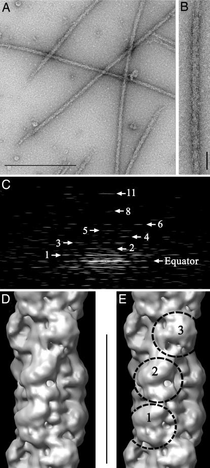
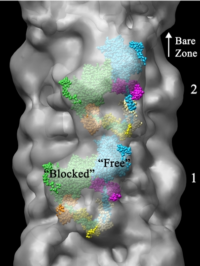
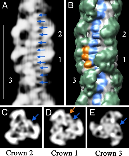
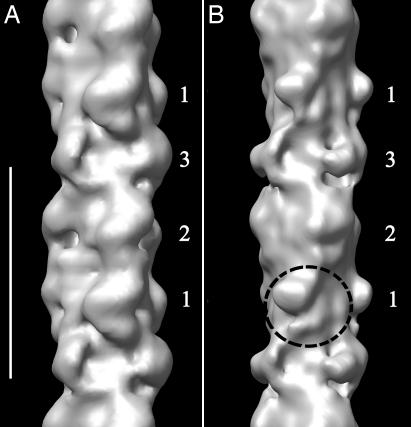
Contraction of the heart results from interaction of the myosin and actin filaments. Cardiac myosin filaments consist of the molecular motor myosin II, the sarcomeric template protein, titin, and the cardiac modulatory protein, myosin binding protein C (MyBP-C). Inherited hypertrophic cardiomyopathy (HCM) is a disease caused mainly by mutations in these proteins. The structure of cardiac myosin filaments and the alterations caused by HCM mutations are unknown. We have used electron microscopy and image analysis to determine the three-dimensional structure of myosin filaments from wild-type mouse cardiac muscle and from a MyBP-C knockout model for HCM. Three-dimensional reconstruction of the wild-type filament reveals the conformation of the myosin heads and the organization of titin and MyBP-C at 4 nm resolution. Myosin heads appear to interact with each other intramolecularly, as in off-state smooth muscle myosin [Wendt T, Taylor D, Trybus KM, Taylor K (2001) Proc Natl Acad Sci USA 98:4361-4366], suggesting that all relaxed muscle myosin IIs may adopt this conformation. Titin domains run in an elongated strand along the filament surface, where they appear to interact with part of MyBP-C and with the myosin backbone. In the knockout filament, some of the myosin head interactions are disrupted, suggesting that MyBP-C is important for normal relaxation of the filament. These observations provide key insights into the role of the myosin filament in cardiac contraction, assembly, and disease. The techniques we have developed should be useful in studying the structural basis of other myosin-related HCM diseases.


Conflict of interest statement

The authors declare no conflict of interest.

Figures

Fig. 1.
Fig. 1.
Isolation and 3D reconstruction of mouse cardiac thick filaments. (A) Electron micrograph of negatively stained filaments. (Scale bar, 500 nm.) (B) Portion of isolated filament, with bare zone at top. (Scale bar, 43 nm.) (C) Fourier transform of B showing layer lines at orders (numbered) of 42.9-nm repeat. (D and E) Surface views of thick filament reconstruction (filtered to 4.0-nm resolution), showing a single 42.9-nm repeat (scale bar), oriented so that bare zone would be at top. Myosin heads project from the surface at three levels (crowns 1, 2, and 3) in each repeat.
Fig. 2.
Fig. 2.
Fitting of atomic model of myosin heads in off-state conformation (24) to head motif in crowns 1 and 2 of reconstruction. The 3D envelope has been made translucent to aid visualization of enclosed atomic model. Small regions of atomic model outside the envelope appear bright, whereas the majority, enclosed within it, appears dull (see also SI Movie 2). The heads are labeled “blocked” and “free” according to terminology proposed to describe the actin-binding capability of each head for regulated myosin (24, 25). “Blocked” head: motor domain, green; essential light chain, orange; regulatory light chain, yellow. “Free” head: motor domain, cyan; essential light chain, pink; regulatory light chain, beige. Bare zone direction toward top.
Fig. 3.
Fig. 3.
Non-myosin proteins in the thick filament. (A) Projection of front side of reconstruction (rear side removed for clarity). Arrows point to 11 densities spaced 4 nm apart within the 42.9-nm repeat (protein white). (Scale bar, 43 nm.) (B) Surface view of same region at higher contour level, showing the three crowns of myosin heads (green). Surface features corresponding to the strand of densities in A are visible (blue, representing titin), together with three additional densities in crown 1 (orange, thought to represent MyBP-C). (C–E) Projections of transverse sections of the central regions of crowns 2, 1, and 3 (where the strand in A runs roughly parallel to the filament axis), showing the radial positions of the densities in A (blue arrows) and of the three additional beads of density in crown 1 in B (orange arrow). Note that, because the filaments have 3-fold rotational symmetry, each of the features marked in A–E is repeated at azimuthal angles of 120° and 240° with respect to those shown.
Fig. 4.
Fig. 4.
Comparison of 3D reconstructions of MyBP-C knockout and wild-type thick filaments. (A) Wild type. (B) Knockout. The reconstructions are based on a similar number of segments in each case. Both have been filtered to 7-nm resolution (the resolution of the knockout filaments) to enable a direct comparison. Circle shows altered structure of myosin heads in crown 1 of knockout. (Scale bar, 43 nm.)

References

    1. Craig R, Woodhead JL. Curr Opin Struct Biol. 2006;16:204–212. - PubMed
    1. Marian AJ. Curr Cardiol Rev. 2005;1:53–63.
    1. Fatkin D, Graham RM. Physiol Rev. 2002;82:945–980. - PubMed
    1. Huxley HE. J Mol Biol. 1963;7:281–308. - PubMed
    1. Kensler RW, Stewart M. J Cell Biol. 1983;96:1797–1802. - PMC - PubMed

Publication types

LinkOut - more resources