. 2008 Feb 6;3(2):e1560.
doi: 10.1371/journal.pone.0001560.

The Plasmodium export element revisited

Affiliations

The Plasmodium export element revisited

Jan Alexander Hiss et al. PLoS One. .

Abstract

We performed a bioinformatical analysis of protein export elements (PEXEL) in the putative proteome of the malaria parasite Plasmodium falciparum. A protein family-specific conservation of physicochemical residue profiles was found for PEXEL-flanking sequence regions. We demonstrate that the family members can be clustered based on the flanking regions only and display characteristic hydrophobicity patterns. This raises the possibility that the flanking regions may contain additional information for a family-specific role of PEXEL. We further show that signal peptide cleavage results in a positional alignment of PEXEL from both proteins with, and without, a signal peptide.


Conflict of interest statement

Competing Interests: The authors have declared that no competing interests exist.

Figures

Figure 1
Figure 1. SOM projection of the PEXEL containing sequence fragments from P.falciparum proteins.
The SOM contains 10×15 topologically ordered data clusters (“neurons”). Locations of RIFIN (green), STEVOR (red), and PfEMP1 (yellow) proteins are highlighted. The location of hypothetical proteins is shown in magenta. Blue color indicates “other” PEXEL-containing fragments. A neuron is assigned one particular class if more than 50% of its clustered proteins belong to one family. White neurons do not contain any proteins (“empty sequence space”). Map generated with the software SOMMER .
Figure 2
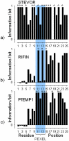
Figure 2. Occurrence of hydrophobic and hydrophilic residues up and downstream the PEXEL motif.
Information plot of the PEXEL motif and surrounding residue positions in the protein families STEVOR (N = 30) (a), RIFIN (N = 125) (b) and PfEMP1 (N = 58) (c). Large values indicate sequence positions with conserved hydrophobic (H) or hydrophilic (O) residues (see text for residue classification). The position of the PEXEL motif is highlighted.
Figure 3
Figure 3. Average position-specific hydrophobicity in 25-residue fragments containing the central PEXEL motif (at positions 11-15).
Color indicates the protein family (black: PfEMP1, gray: RIFIN, light gray: STEVOR). Error bars give standard deviations. Asterisks indicate positions characteristic for one of the families. Note that the Hopp & Woods scale is a “hydrophilicity” scale with negative values for hydrophobic residues.
Figure 4
Figure 4. Distribution of the first position of the PEXEL motif.
A) position of the PEXEL motif in sequences of the PfEMP1 protein family (red), exported proteins with a predicted signal peptide (blue), and exported proteins lacking a predicted signal peptide (gray). In B) the blue bars show the positions of the PEXEL motif after cleaving off the predicted signal peptide. Only sequences with a predicted signal peptidase cleavage site (score >0.5 according to SignalP [38], [39]) are included. Gray bars in B) represent the unchanged distribution of PEXEL in proteins lacking a predicted signal sequence. Note that all bars are displayed on top of each other.

References

    1. Cooke BM, Lingelbach K, Bannister LH, Tilley L. Protein trafficking in Plasmodium falciparum-infected red blood cells. Trends Parasitol. 2004;20:581–589. - PubMed
    1. Hiller NL, Bhattacharjee S, van Ooij C, Liolios K, Harrison T, et al. A host-targeting signal in virulence proteins reveals a secretome in malarial infection. Science. 2004;306:1934–1937. - PubMed
    1. Marti M, Good RT, Rug M, Knuepfer E, Cowman AF. Targeting malaria virulence and remodeling proteins to the host erythrocyte. Science. 2004;306:1930–1933. - PubMed
    1. Smith JD, Chitnis CE, Craig AG, Roberts DJ, Hudson-Taylor DE, et al. Switches in expression of Plasmodium falciparum var genes correlate with changes in antigenic and cytoadherent phenotypes of infected erythrocytes. Cell. 1995;82:101–110. - PMC - PubMed
    1. Baruch DI, Pasloske BL, Singh HB, Bi X, Ma XC, et al. Cloning the P. falciparum gene encoding PfEMP1, a malarial variant antigen and adherence receptor on the surface of parasitized human erythrocytes. Cell. 1995;82:77–87. - PubMed

Publication types