Angiography underestimates peripheral atherosclerosis: lumenography revisited
- PMID: 18254670
- PMCID: PMC2795386
- DOI: 10.1583/07-2249R.1
Angiography underestimates peripheral atherosclerosis: lumenography revisited
Abstract
Purpose: To compare angiograms, considered the gold standard for diagnostic imaging of peripheral arterial disease (PAD), to the corresponding histological sections of popliteal and tibial vessels obtained after amputation to determine if angiography fails to define atheroma burden in "normal appearing" arteries in patients with PAD.
Methods: Between 2004 and 2006, 69 patients underwent amputation of a lower extremity for severe tissue loss, gangrene, or pedal sepsis precluding limb salvage. Popliteal and tibial vessels were harvested, perfusion-fixed, and analyzed histologically. Thirty-four of these patients had pre-amputation angiography during attempted salvage procedures. Angiograms with patent or minimally diseased vessel segments (n = 19) were assessed for stenoses, diameter, and calcification by 3 vascular surgeons (n = 72 evaluations). These results were compared to corresponding cross-sectional histological slides (n = 66) in a blinded manner.
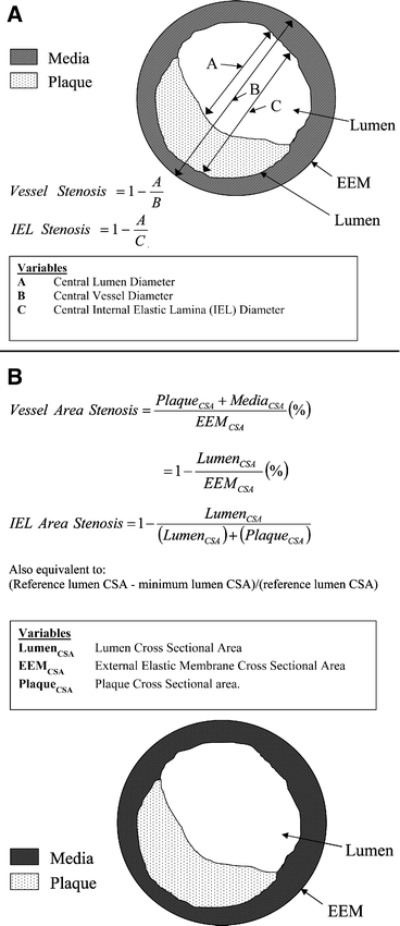
Results: Angiograms performed prior to above-knee (n = 9) or below-knee (n = 10) amputation revealed 24 stenoses with a mean (+/-SD) diameter-reducing stenosis of 19.5%+/-15.2%. Corresponding histological cross sections revealed greater linear stenoses measured via boundaries of the internal elastic lamina (IEL stenosis, 28.9%+/-20.2%, p = 0.003 versus angiography) or via boundaries of the external elastic membrane (vessel stenosis, 43.1%+/-15.2%, p<0.0001). Stenosis calculated by area methods (IEL area) were greater and measured 39.2%+/-24.2% (p<0.0001) and 60.9%+/-15.2% (vessel area, p<0.0001). Popliteal arteries had greater discrepancy in stenosis measurement than tibial arteries (18.5%+/-14.6% versus 34.9%+/-21.0%, p = 0.0005). However, evaluations of tibial arteries for concentricity of plaque (44% versus 69%, p = 0.08) and calcification grade (1.6 versus 2.2, p = 0.002) by angiography were discordant with histological analyses. Measurement of arterial diameter by histology for popliteal arteries (6.2+/-0.9 mm) and tibial arteries (3.1+/-0.7 mm) was greater than angiographic diameter determination (p<0.001).
Conclusion: Angiography provides information on luminal characteristics of peripheral arteries but severely underestimates the extent of atherosclerosis in patients with PAD even in "normal appearing" vessels.
Figures


References
-
- Sones F.M., Shirey E.K. Cine coronary arteriography. Mod Concepts Cardiovasc Dis. 1962;31:735–738. - PubMed
-
- Kruger R.A., Mistretta C.A., Crummy A.B. et al.Digital K-edge subtraction radiography. Radiology. 1977;125:243–245. - PubMed
-
- Dotter C.T., Judkins M.P. Transluminal treatment of arteriosclerotic obstruction. Description of a new technic and a preliminary report of its application. Circulation. 1964;30:654–670. - PubMed
-
- Kronenberg F., Mundle M., Langle M. et al.Prevalence and progression of peripheral arterial calcifications in patients with ESRD. Am J Kidney Dis. 2003;41:140–148. - PubMed
-
- Vlodaver Z., Frech R., Van Tassel R.A. et al.Correlation of the antemortem coronary arteriogram and the postmortem specimen. Circulation. 1973;47:162–169. - PubMed
Publication types
MeSH terms
Grants and funding
LinkOut - more resources
Full Text Sources
Other Literature Sources
Medical

