. 2008 Feb 6:8:41.
doi: 10.1186/1471-2148-8-41.

Identification of the Otopetrin Domain, a conserved domain in vertebrate otopetrins and invertebrate otopetrin-like family members

Affiliations

Identification of the Otopetrin Domain, a conserved domain in vertebrate otopetrins and invertebrate otopetrin-like family members

Inna Hughes et al. BMC Evol Biol. .

Abstract

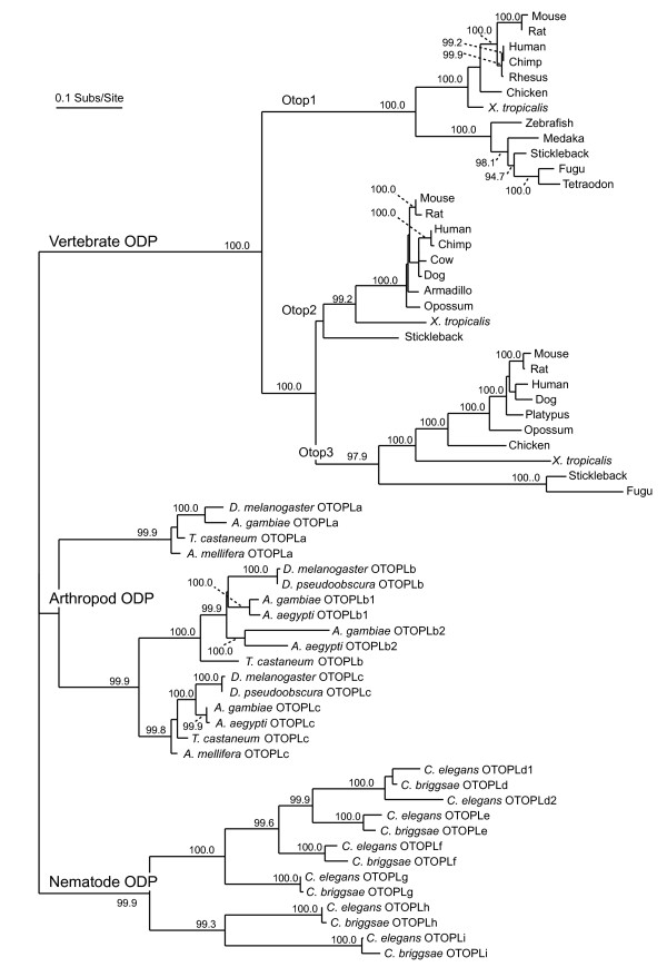
Background: Otopetrin 1 (Otop1) encodes a multi-transmembrane domain protein with no homology to known transporters, channels, exchangers, or receptors. Otop1 is necessary for the formation of otoconia and otoliths, calcium carbonate biominerals within the inner ear of mammals and teleost fish that are required for the detection of linear acceleration and gravity. Vertebrate Otop1 and its paralogues Otop2 and Otop3 define a new gene family with homology to the invertebrate Domain of Unknown Function 270 genes (DUF270; pfam03189).

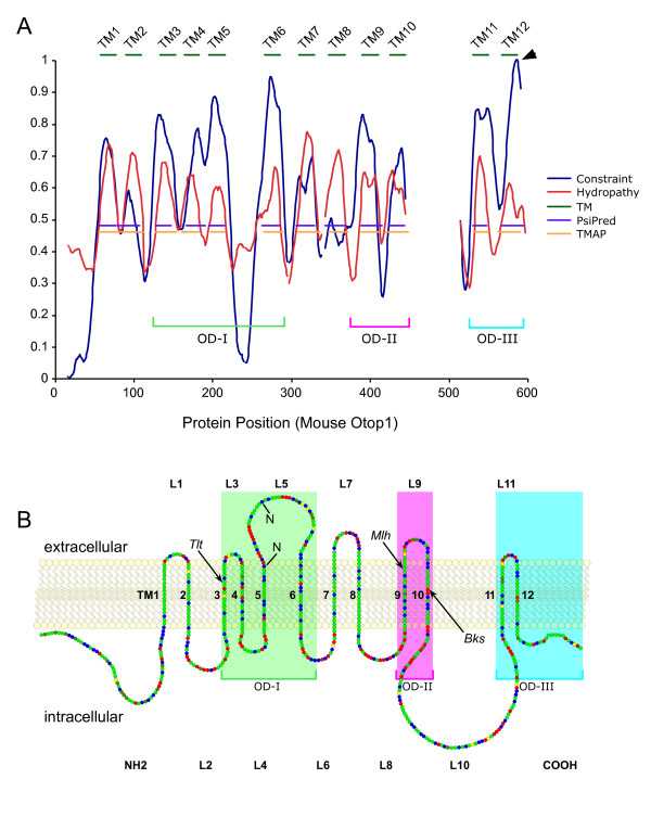
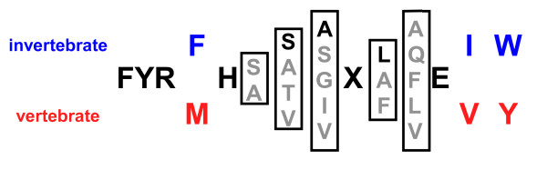
Results: Multi-species comparison of the predicted primary sequences and predicted secondary structures of 62 vertebrate otopetrin, and arthropod and nematode DUF270 proteins, has established that the genes encoding these proteins constitute a single family that we renamed the Otopetrin Domain Protein (ODP) gene family. Signature features of ODP proteins are three "Otopetrin Domains" that are highly conserved between vertebrates, arthropods and nematodes, and a highly constrained predicted loop structure.

Conclusion: Our studies suggest a refined topologic model for ODP insertion into the lipid bilayer of 12 transmembrane domains, and highlight conserved amino-acid residues that will aid in the biochemical examination of ODP family function. The high degree of sequence and structural similarity of the ODP proteins may suggest a conserved role in the intracellular trafficking of calcium and the formation of biominerals.


Figures

Figure 1
Figure 1
Phylogeny of the Otopetrin Domain Protein (ODP) family. Maximum-likelihood phylogenetic tree created from the multi-sequence alignment of 62 ODPs (see additional file 1). The vertebrate, arthropod, and nematode sequences form distinct monophyletic groups, each containing three or more paralogous groups. Some nematode and arthropod sequences appear to have undergone additional gene-duplication events, creating species-specific paralogs (designated with a 1 or 2 following the gene symbol). Branch labels are bootstrap values for 1000 replicates. Unlabeled internal branches have bootstrap values less than 90.0.
Figure 2
Figure 2
Predicted secondary structure and topologic model for Otop1 insertion into the lipid bilayer. A) Hydrophobicity (red) and evolutionary constraint (blue) are plotted against the amino-acid position of mouse Otop1. A total of 12 evolutionarily constrained regions are found in the ODP family that are highly hydrophobic and have a helical structure consistent with TM domains (dark green), as predicted by TMAP (orange) and PsiPred (purple). Green, pink, and blue brackets define the highly conserved subdomains: Otopetrin Domain-I, -II, and -III (OD-I, OD-II, and OD-III, respectively). B) Linear model of mouse Otop1a inserted in a lipid bilayer, in which each amino acid is represented as a circle and the chemical properties of amino-acids are denoted by color: charged residues (red), polar residues (blue), and non-polar residue (green). Cysteine (yellow) and proline (dark green) are noted. The two consensus N-glycosylation sites (N) are indicated in loop 5. The predicted intracellular and extracellular loops and TM domains are numbered L1 to L11 and TM1 to TM12, respectively. The locations of the tlt, mlh, and bks mutations are noted by arrows. The three OD subdomains are shaded with the color code used in A.
Figure 3
Figure 3
FYR box consensus sequence for the ODP family C-terminal tail. Residues in bold are shared by all ODP family members, X is any hydrophobic amino acid, blue residues are specifically conserved in arthropod and nematode members, and red amino acids are conserved among vertebrate members. Grey, bracketed residues represent common variants at each less-conserved position. The dark residue within each bracket represents the most common amino-acid variant at that position, if one can be identified.

References

    1. Gizzi M, Ayyagari S, Khattar V. The familial incidence of benign paroxysmal positional vertigo. Acta Otolaryngol. 1998;118(6):774–777. doi: 10.1080/00016489850182422. - DOI - PubMed
    1. Oghalai JS, Manolidis S, Barth JL, Stewart MG, Jenkins HA. Unrecognized benign paroxysmal positional vertigo in elderly patients. Otolaryngol Head Neck Surg. 2000;122(5):630–634. doi: 10.1016/S0194-5998(00)70187-2. - DOI - PubMed
    1. Oas JG. Benign paroxysmal positional vertigo: a clinician's perspective. Ann N Y Acad Sci. 2001;942:201–209. - PubMed
    1. Tusa RJ. Benign paroxysmal positional vertigo. Curr Neurol Neurosci Rep. 2001;1(5):478–485. doi: 10.1007/s11910-001-0110-y. - DOI - PubMed
    1. Bronstein AM. Benign paroxysmal positional vertigo: some recent advances. Curr Opin Neurol. 2003;16(1):1–3. doi: 10.1097/00019052-200302000-00001. - DOI - PubMed

Publication types

MeSH terms

Substances

LinkOut - more resources