. 2008 Apr;29(12):1976-88.
doi: 10.1016/j.biomaterials.2007.12.033. Epub 2008 Feb 6.

Dynamic imaging of arginine-rich heart-targeted vehicles in a mouse model

Affiliations

Dynamic imaging of arginine-rich heart-targeted vehicles in a mouse model

Hua Zhang et al. Biomaterials. 2008 Apr.

Abstract

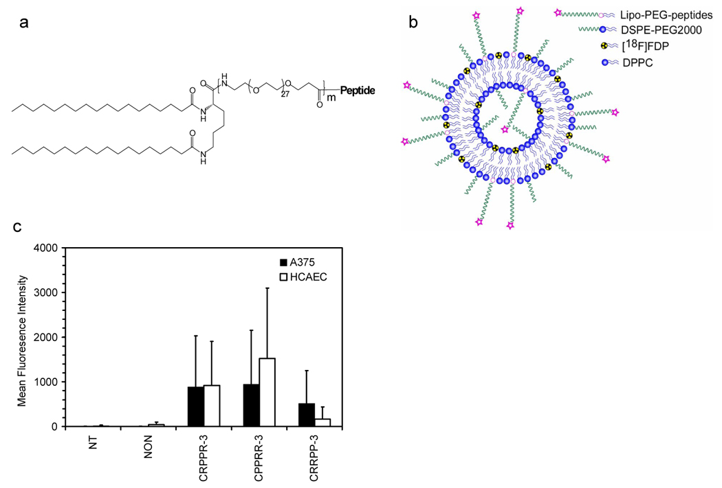
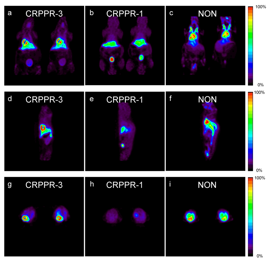
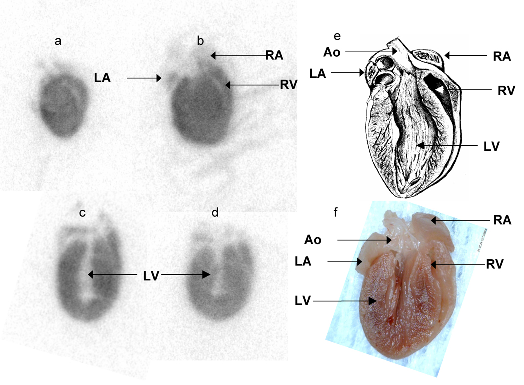
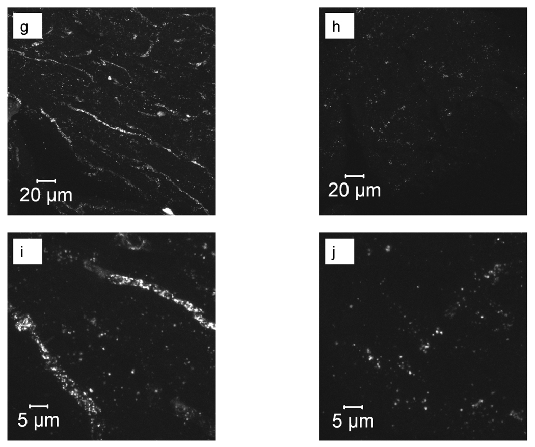
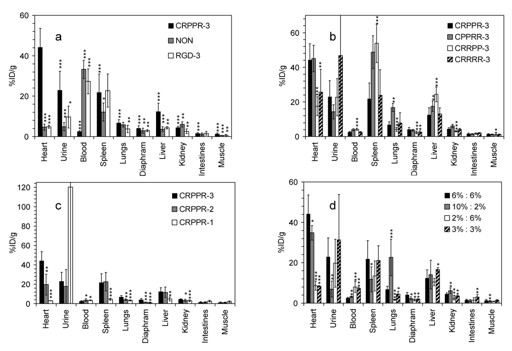
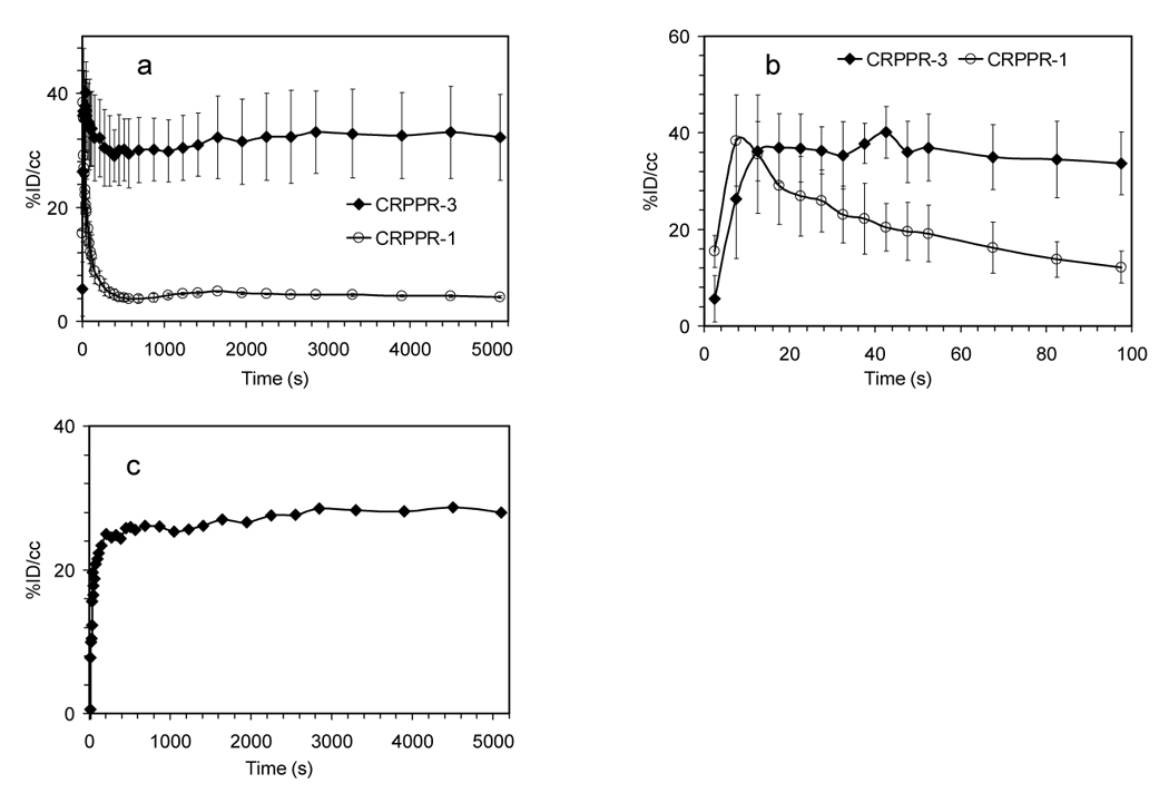
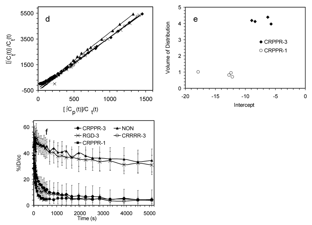
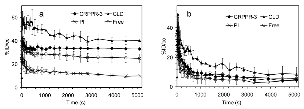
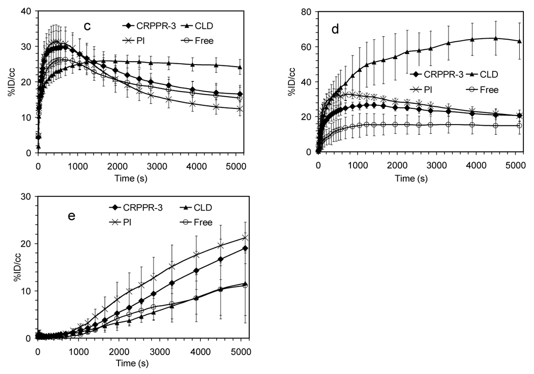
Efficacious delivery of drugs and genes to the heart is an important goal. Here, a radiolabeled peptide-targeted liposome was engineered to bind to the heart, and the biodistribution and pharmacokinetics were determined by dynamic positron emission tomography in the FVB mouse. Efficient targeting occurred only with an exposed ligand and a dense concentration of peptide (6000 peptides/particles). Liposomes targeted with CRPPR or other arginine-rich peptides with an exposed guanidine moiety bound within 100 s after intravenous injection and remained stably bound. With CRPPR-targeted particles, the radioisotope density in the heart averaged 44 +/- 9% injected dose/gram of tissue, more than 30-fold higher than in skeletal muscle. The rapid and efficient targeting of these particles can be exploited in drug and gene delivery systems and with dynamic positron emission tomography provides a model system to optimize targeting of engineered particles.


Figures

Figure 1
Figure 1
(a) Chemical structures of lipo-PEG-peptides (LPPs) and (b) schematic of radiolabeled targeted liposome. Molecular weights of the PEG spacer are 1200, 2400, 3600 for m =1, 2, 3, respectively. Abbreviations for the LPPs are as follows. CRPPR-3: peptide = CRPPR, m = 3; CRPPR-2: peptide = CRPPR, m = 2; CRPPR-1: peptide = CRPPR, m = 1; RGD-3: peptide = c(RGDY(OMe)KE), m = 3; CPPRR-3: peptide = CPPRR, m = 3; CRRPP-3: peptide = CRRPP, m = 3; CRRRR-3: peptide = CRRRR, m = 3; NON indicates no LPP (but 12% DSPE-PEG2000); NT indicates in vitro incubation without liposomes. LPPs are incorporated within a liposome prior to injection with a formulation of LPP : DSPE-PEG2000 : DPPC = 6:6:88 (mol/mol), except in figure 4d. (c) Fluorescence intensity, as measured by flow cytometry, for a melanoma cell line, A375, and an endothelial cell line, Human Coronary Artery Endothelial Cells (HCAEC) incubated with liposomes containing CRPPR-3 lipo-PEG-peptides. Fluorescent, viable cells were quantified after incubation and washing.
Figure 2
Figure 2
(a–i) 90-minute accumulative PET images acquired after injection of radiolabeled liposomes from coronal (a–c), sagittal (d–f) and transverse views (g–i) with LPPs CRPPR-3 (a,d,g), CRPPR-1 (b,e,h), and NON (c,f,i).
Figure 3
Figure 3
High resolution autoradiography and optical imaging of the heart after injection of targeted and control particles. (a–d) Autoradiography images acquired from 60 µm tissue slices 90 minutes after injection of CRPPR-3 liposomes. (e) Anatomic drawing of a mouse heart with the same orientation as the processed tissue slices (picture by William Moroski). (f) Digital photograph of a mouse heart fixed in 10% formalin. A for atrium, V for ventricle, L for left, R for right and Ao for aorta. (g–j) Confocal microscopy images of heart tissue after intravenous injection of CRPPR-3- (g, i), and NON- (h, j) targeted liposomes with low (g, h) and high (i, j) magnification.
Figure 3
Figure 3
High resolution autoradiography and optical imaging of the heart after injection of targeted and control particles. (a–d) Autoradiography images acquired from 60 µm tissue slices 90 minutes after injection of CRPPR-3 liposomes. (e) Anatomic drawing of a mouse heart with the same orientation as the processed tissue slices (picture by William Moroski). (f) Digital photograph of a mouse heart fixed in 10% formalin. A for atrium, V for ventricle, L for left, R for right and Ao for aorta. (g–j) Confocal microscopy images of heart tissue after intravenous injection of CRPPR-3- (g, i), and NON- (h, j) targeted liposomes with low (g, h) and high (i, j) magnification.
Figure 4
Figure 4
Well counts (%ID/g) obtained 90 minutes after injection. (a), (b) and (c), with different LPP; (d), with varied ratios of CRPPR-3:DSPE-PEG2000. Significance of the accumulation of particles targeted with CRPPR-3:DSPE-PEG2000 6%:6% tested against other peptides and surface architectures is shown by ***, p<0.001; ** , p<0.01; *, p<0.05. In 4a with CRPPR-3, accumulation in each organ is tested against accumulation in the heart. Significance of accumulation is otherwise tested against CRPPR-3 in the same organ.
Figure 5
Figure 5
Time activity curves (TACs) and Logan Plot from dynamic PET analysis of various LPP liposomes. (a)–(b) TACs for heart muscle, (c) difference between CRPPR-3 and CRPPR-1 liposomes in TAC from heart muscle. Subtraction of non-binding CRPPR-1 particles removes the effect of the blood pool, showing accumulation of particles within heart muscle. (d) results of Logan analysis for 4 injections of CRPPR-3 liposomes, plotting the time integral of activity at target, Ct(t) against the integral of activity in blood, Cp(t), each normalized by Ct(t). (e) summary of slope and intercept for plots as shown in (d). The higher volume of distribution of CRPPR-3 particles indicates higher avidity. (f – i) TACs for regions of interest. (f) blood within the heart chamber, (g) liver, (h) spleen, and (i) bladder.
Figure 5
Figure 5
Time activity curves (TACs) and Logan Plot from dynamic PET analysis of various LPP liposomes. (a)–(b) TACs for heart muscle, (c) difference between CRPPR-3 and CRPPR-1 liposomes in TAC from heart muscle. Subtraction of non-binding CRPPR-1 particles removes the effect of the blood pool, showing accumulation of particles within heart muscle. (d) results of Logan analysis for 4 injections of CRPPR-3 liposomes, plotting the time integral of activity at target, Ct(t) against the integral of activity in blood, Cp(t), each normalized by Ct(t). (e) summary of slope and intercept for plots as shown in (d). The higher volume of distribution of CRPPR-3 particles indicates higher avidity. (f – i) TACs for regions of interest. (f) blood within the heart chamber, (g) liver, (h) spleen, and (i) bladder.
Figure 6
Figure 6
TACs from dynamic PET analysis for (a) heart muscle (b) blood pool (c) liver (d) spleen (e) bladder after injection of CRPPR-3 liposomes and various inhibitors. Abbreviations: CRPPR-3: no inhibitors; CLD: clodronate liposomes injected 24 hours in advance; PI: poly(inosinic acid) injected 1 minute in advance; Free: free CRPPR peptide injected 1 minute in advance of CRPPR-3 liposome injection. Well counts at 90 minutes after injection: the ratios of radioactivity in the heart with and without inhibitors are 1.30, 0.59 and 0.81 for CLD, PI and Free, p = 0.10, < 0.01, and < 0.05, respectively.
Figure 6
Figure 6
TACs from dynamic PET analysis for (a) heart muscle (b) blood pool (c) liver (d) spleen (e) bladder after injection of CRPPR-3 liposomes and various inhibitors. Abbreviations: CRPPR-3: no inhibitors; CLD: clodronate liposomes injected 24 hours in advance; PI: poly(inosinic acid) injected 1 minute in advance; Free: free CRPPR peptide injected 1 minute in advance of CRPPR-3 liposome injection. Well counts at 90 minutes after injection: the ratios of radioactivity in the heart with and without inhibitors are 1.30, 0.59 and 0.81 for CLD, PI and Free, p = 0.10, < 0.01, and < 0.05, respectively.

Similar articles

Cited by

References

    1. Ding BS, Dziubla T, Shuvaev VV, Muro S, Muzykantov VR. Advanced drug delivery systems that target the vascular endothelium. Molecular Interventions. 2006;6(2):98–112. - PubMed
    1. Hajitou A, Pasqualini R, Arap W. Vascular targeting: Recent advances and therapeutic perspectives. Trends in Cardiovascular Medicine. 2006;16(3):80–88. - PMC - PubMed
    1. Ruoslahti E. Vascular zip codes in angiogenesis and metastasis. Biochem Soc Trans. 2004;32:397–402. - PubMed
    1. Zhang LL, Hoffman JA, Ruoslahti E. Molecular profiling of heart endothelial cells. Circulation. 2005;112(11):1601–1611. - PubMed
    1. Brissette R, Prendergast JKA, Goldstein NI. Identification of cancer targets and therapeutics using phage display. Curr Opin Drug Discov Dev. 2006;9(3):363–369. - PubMed

Publication types

MeSH terms