. 2008 Apr 18;283(16):10872-80.
doi: 10.1074/jbc.M708719200. Epub 2008 Feb 6.

Allosteric motions in structures of yeast NAD+-specific isocitrate dehydrogenase

Affiliations

Allosteric motions in structures of yeast NAD+-specific isocitrate dehydrogenase

Alexander B Taylor et al. J Biol Chem. .

Abstract

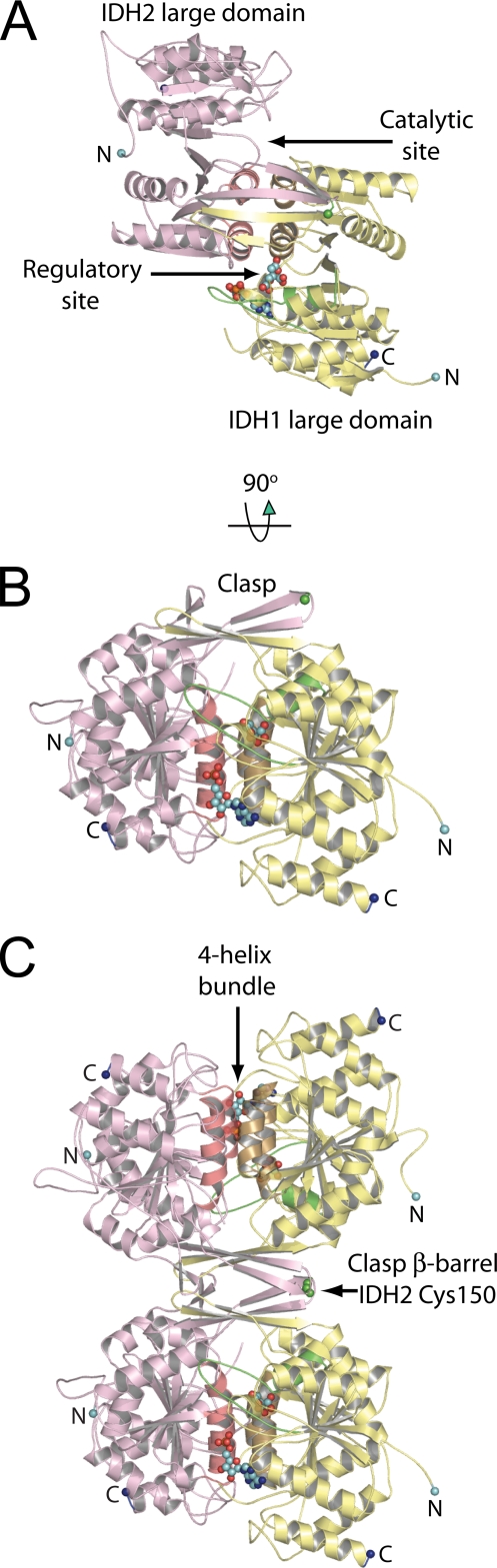
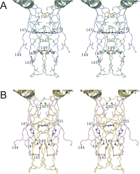
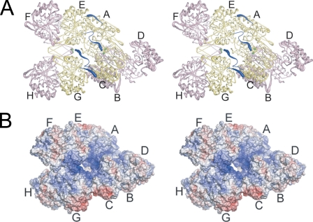
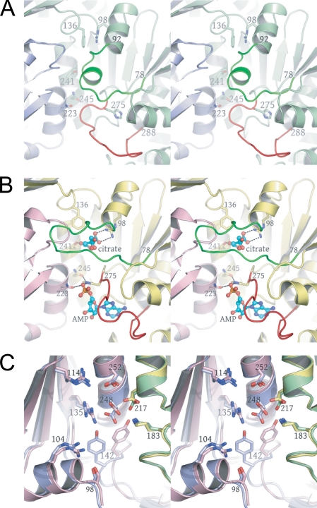
Mitochondrial NAD(+)-specific isocitrate dehydrogenases (IDHs) are key regulators of flux through biosynthetic and oxidative pathways in response to cellular energy levels. Here we present the first structures of a eukaryotic member of this enzyme family, the allosteric, hetero-octameric, NAD(+)-specific IDH from yeast in three forms: 1) without ligands, 2) with bound analog citrate, and 3) with bound citrate + AMP. The structures reveal the molecular basis for ligand binding to homologous but distinct regulatory and catalytic sites positioned at the interfaces between IDH1 and IDH2 subunits and define pathways of communication between heterodimers and heterotetramers in the hetero-octamer. Disulfide bonds observed at the heterotetrameric interfaces in the unliganded IDH hetero-octamer are reduced in the ligand-bound forms, suggesting a redox regulatory mechanism that may be analogous to the "on-off" regulation of non-allosteric bacterial IDHs via phosphorylation. The results strongly suggest that eukaryotic IDH enzymes are exquisitely tuned to ensure that allosteric activation occurs only when concentrations of isocitrate are elevated.


Figures

FIGURE 1.
FIGURE 1.
The yeast IDH heterodimer and heterotetramer. The regulatory IDH1 subunits are shown in yellow and the catalytic IDH2 subunits in pink. The N and C termini are shown as cyan and dark blue spheres, respectively. Helices g and h, which form the four-helix bundle at the heterodimer interface, are shown in gold and salmon coming from IDH1 and IDH2, respectively. Citrate and AMP are shown as spheres bound to IDH1. Residues 78-92 of IDH1, which undergo the helix-loop structural transition upon ligand binding, are shown in green. A, the IDH heterodimer shown looking down the pseudo-2-fold axis of rotation. The regulatory and catalytic sites and the large domains in each subunit are labeled. B, the IDH heterodimer shown looking perpendicular to the pseudo-2-fold axis of rotation. The β-hairpins formed by β-strands K and L and comprising the clasp region are indicated. The sulfur atom of Cys-150 coming from IDH2 in the turn between β-strands K and L is shown as a green sphere. C, the yeast IDH heterotetramer. The lower heterodimer is shown in the same orientation as in B. The 2-fold axis of rotation formed at the clasp β-barrel heterotetrameric interface is horizontal in the plane of the page. The disulfide bond formed in the unliganded IDH structure by IDH2 Cys-150 residues is indicated.
FIGURE 2.
FIGURE 2.
Alterations at the clasp β-barrel formed at the heterotetrameric interface in the unliganded and citrate-bound yeast IDH structures. The interior of the barrel is formed by alternating layers of apolar and polar residues (see “Results”). Hydrogen bonds formed by polar side chains are shown as dotted lines. A, the clasp β-barrel in the unliganded yeast IDH structure. IDH1 and IDH2 subunits are shown in green and slate blue, respectively. IDH2 Cys-150 residues form a disulfide bond (bright yellow). The five-turn helices formed by IDH1 residues 157-174 are positioned such that the uncapped N termini are adjacent to the Cys-150 Sγ atoms, which are centered on the long helix axes (see “Results” and supplemental Figs. 6 and 7). B, the clasp β-barrel in the citrate-bound yeast IDH structure shown in the same orientation as in A. IDH1 and IDH2 subunits are shown in yellow and pink, respectively. The disulfide bond between IDH2 Cys-150 residues (bright green) is reduced, the side chain of IDH2 His-147 disrupts the lower adjacent apolar layer of side chains, and the remaining polar side chains form fewer hydrogen bonds than in the unliganded IDH structure.
FIGURE 3.
FIGURE 3.
The yeast IDH hetero-octamer does not obey pseudo-222 symmetry. Regulatory IDH1 subunits are shown in yellow and catalytic IDH2 subunits in pink. The N termini of IDH1 subunits are shown in dark blue, and the Sγ atoms of IDH2 Cys-150 residues are shown as bright green spheres. The orientation of the leftmost heterotetramer in each panel is in the same orientation as in Fig. 1C. The thickness of the tubes is proportional to the thermal parameters for the backbone atoms, with thicker tubes representing higher atomic displacement parameters. A, the yeast IDH hetero-octamer with citrate bound. Note that the N termini of two IDH1 subunits run along the interior of the central solvent-filled cavity of the hetero-octamer and the remaining two IDH1 N termini are positioned on the exterior of the hetero-octamer. The interior IDH1 N termini from each heterotetramer interact reciprocally with the Cys-150 residues of theβ-barrel clasp of the other heterotetramer in the ligand-bound but not in the unliganded IDH structures (see “Results”). The 2-fold axis of rotation relating heterotetramers runs normal to a line running from ∼1:00 to 7:00 o'clock in the plane of the page. B, electrostatic surface potential calculated at ±10 kT for the citrate-bound yeast IDH hetero-octamer. The orientation is the same as shown in panel A and in Fig. 1C. Basic amino acid side chains coming from the uncapped IDH1 helices consisting of residues 156-173 and the proximity of the N-terminal amino group of IDH1 chain C and E contribute to a substantial positive electrostatic potential (blue shading) at IDH2 residues Cys-150 (see “Results”).
FIGURE 4.
FIGURE 4.
The regulatory and catalytic sites in the unliganded (green and slate blue) and citrate-bound (yellow and pink) yeast IDH heterodimers. Amino acid residues undergoing a conformational change upon citrate binding are shown in bright green and red. Citrate and AMP are shown as ball-and-stick, and the side chains of residues involved in ligand binding are shown as sticks alone. The AMP-binding site was identified in difference electron density maps after rigid body refinement of the citrate-bound IDH structure against the 4.3 Å citrate + AMP diffraction data (see “Experimental Procedures” and Fig. 5A). A, regulatory IDH1-binding site in the unliganded IDH structure. Note the position of the green helix corresponding to residues 78-92 and the large red loop corresponding to residues 275-285. These residues block both the citrate- and AMP-binding sites, respectively. B, regulatory IDH1-binding site in the citrate-bound IDH structure. Upon citrate binding, the green residues undergo a helix-loop transition that causes a shift in the position of the red loop that in turn opens the AMP-binding site (see “Results”). C, catalytic IDH2-binding sites with the same color coding as shown in A. The side chains of catalytically invariant Arg-104, Arg-114, Arg-135, Tyr-142, Lys-183, Asp-217, Asp-248, and Asp-252 are shown as ball-and stick.
FIGURE 5.
FIGURE 5.
Difference and annealed omit electron density superimposed on key regions of IDH. A, the AMP-binding site is revealed by Fo - Fc electron density contoured at 3σ after rigid body refinement of the citrate-bound IDH structure against the 4.3 Å citrate + AMP diffraction data. The orientation is approximately the same as that shown in Fig. 4, A and B. B, annealed omit map contoured at 3σ calculated for the unliganded IDH structure in which IDH2 residues 148-153 were removed from the phase calculations. The electron density reveals an oxidized disulfide bond between symmetry-related IDH2 Cys-150 residues. C, annealed omit map contoured at 4σ calculated for the citrate-bound IDH structure in which IDH2 residues 148-153 were removed from the phase calculations. The electron density reveals a reduced disulfide bond between symmetry-related IDH2 Cys-150 residues (see “Results”).

References

    1. Chen, R. F., and Plaut, G. W. (1963) Biochemistry 2 1023-1032 - PubMed
    1. Plaut, G. W. (1970) Curr. Top. Cell. Regul. 2 1-27
    1. Barnes, L. D., McGuire, J. J., and Atkinson, D. E. (1972) Biochemistry 11 4322-4329 - PubMed
    1. Gabriel, J. L., Zervos, P. R., and Plaut, G. W. (1986) Metabolism 35 661-667 - PubMed
    1. Sols, A., Gancedo, C., and DelaFuente, G. (1971) in The Yeasts, (Rose, A. H., and Harrison, J. S., eds) 1st Ed., Vol. 2, pp. 271-307, Academic Press, London

Publication types

Associated data

LinkOut - more resources