hAda3 degradation by papillomavirus type 16 E6 correlates with abrogation of the p14ARF-p53 pathway and efficient immortalization of human mammary epithelial cells
- PMID: 18256148
- PMCID: PMC2293003
- DOI: 10.1128/JVI.02466-07
hAda3 degradation by papillomavirus type 16 E6 correlates with abrogation of the p14ARF-p53 pathway and efficient immortalization of human mammary epithelial cells
Abstract
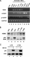
Two activities of human papillomavirus type 16 E6 (HPV16 E6) are proposed to contribute to the efficient immortalization of human epithelial cells: the degradation of p53 protein and the induction of telomerase. However, the requirement for p53 inactivation has been debated. Another E6 target is the hAda3 protein, a p53 coactivator and a component of histone acetyltransferase complexes. We have previously described the role of hAda3 and p53 acetylation in p14ARF-induced human mammary epithelial cell (MEC) senescence (P. Sekaric, V. A. Shamanin, J. Luo, and E. J. Androphy, Oncogene 26:6261-6268, 2007). In this study, we analyzed a set of HPV16 E6 mutants for the ability to induce hAda3 degradation. E6 mutants that degrade hAda3 but not p53 could abrogate p14ARF-induced growth arrest despite the presence of normal levels of p53 and efficiently immortalized MECs. However, two E6 mutants that previously were reported to immortalize MECs with low efficiency were found to be defective for both p53 and hAda3 degradation. We found that these immortal MECs select for reduced p53 protein levels through a proteasome-dependent mechanism. The findings strongly imply that the inactivation of the p14ARF-p53 pathway, either by the E6-mediated degradation of p53 or hAda3 or by cellular adaptation, is required for MEC immortalization.
Figures


Similar articles
-
hAda3 regulates p14ARF-induced p53 acetylation and senescence.Oncogene. 2007 Sep 20;26(43):6261-8. doi: 10.1038/sj.onc.1210462. Epub 2007 Apr 23. Oncogene. 2007. PMID: 17452980
-
Mutational analysis of human papillomavirus type 16 E6 demonstrates that p53 degradation is necessary for immortalization of mammary epithelial cells.J Virol. 1996 Feb;70(2):683-8. doi: 10.1128/JVI.70.2.683-688.1996. J Virol. 1996. PMID: 8551603 Free PMC article.
-
Immortalization of human mammary epithelial cells is associated with inactivation of the p14ARF-p53 pathway.Mol Cell Biol. 2004 Mar;24(5):2144-52. doi: 10.1128/MCB.24.5.2144-2152.2004. Mol Cell Biol. 2004. PMID: 14966292 Free PMC article.
-
Multiple functions of human papillomavirus type 16 E6 contribute to the immortalization of mammary epithelial cells.J Virol. 1999 Sep;73(9):7297-307. doi: 10.1128/JVI.73.9.7297-7307.1999. J Virol. 1999. PMID: 10438818 Free PMC article.
-
Human papillomavirus oncoprotein E6 inactivates the transcriptional coactivator human ADA3.Mol Cell Biol. 2002 Aug;22(16):5801-12. doi: 10.1128/MCB.22.16.5801-5812.2002. Mol Cell Biol. 2002. PMID: 12138191 Free PMC article.
Cited by
-
Human papillomavirus 16 oncoprotein E7 stimulates UBF1-mediated rDNA gene transcription, inhibiting a p53-independent activity of p14ARF.PLoS One. 2014 May 5;9(5):e96136. doi: 10.1371/journal.pone.0096136. eCollection 2014. PLoS One. 2014. PMID: 24798431 Free PMC article.
-
Molecular Probing of the HPV-16 E6 Protein Alpha Helix Binding Groove with Small Molecule Inhibitors.PLoS One. 2016 Feb 25;11(2):e0149845. doi: 10.1371/journal.pone.0149845. eCollection 2016. PLoS One. 2016. PMID: 26915086 Free PMC article.
-
Genomic instability: Ada3 and HPV E6-acetyltransferase connections?Cell Cycle. 2013 Jan 1;12(1):13. doi: 10.4161/cc.23172. Epub 2012 Dec 19. Cell Cycle. 2013. PMID: 23255094 Free PMC article. No abstract available.
-
Tale of a multifaceted co-activator, hADA3: from embryogenesis to cancer and beyond.Open Biol. 2016 Sep;6(9):160153. doi: 10.1098/rsob.160153. Open Biol. 2016. PMID: 27605378 Free PMC article. Review.
-
Virology and molecular pathogenesis of HPV (human papillomavirus)-associated oropharyngeal squamous cell carcinoma.Biochem J. 2012 Apr 15;443(2):339-53. doi: 10.1042/BJ20112017. Biochem J. 2012. PMID: 22452816 Free PMC article. Review.
References
-
- Balasubramanian, R., M. G. Pray-Grant, W. Selleck, P. A. Grant, and S. Tan. 2002. Role of the Ada2 and Ada3 transcriptional coactivators in histone acetylation. J. Biol. Chem. 2777989-7995. - PubMed
-
- Brooks, C. L., and W. Gu. 2003. Ubiquitination, phosphorylation and acetylation: the molecular basis for p53 regulation. Curr. Opin. Cell Biol. 15164-171. - PubMed
-
- Caldeira, S., R. Filotico, R. Accardi, I. Zehbe, S. Franceschi, and M. Tommasino. 2004. p53 mutations are common in human papillomavirus type 38-positive non-melanoma skin cancers. Cancer Lett. 209119-124. - PubMed
Publication types
MeSH terms
Substances
Grants and funding
LinkOut - more resources
Full Text Sources
Other Literature Sources
Research Materials
Miscellaneous

