. 2008 Mar;135(6):1147-56.
doi: 10.1242/dev.018150. Epub 2008 Feb 6.

The ATPase-dependent chaperoning activity of Hsp90a regulates thick filament formation and integration during skeletal muscle myofibrillogenesis

Affiliations

The ATPase-dependent chaperoning activity of Hsp90a regulates thick filament formation and integration during skeletal muscle myofibrillogenesis

Thomas A Hawkins et al. Development. 2008 Mar.

Abstract

The mechanisms that regulate sarcomere assembly during myofibril formation are poorly understood. In this study, we characterise the zebrafish sloth(u45) mutant, in which the initial steps in sarcomere assembly take place, but thick filaments are absent and filamentous I-Z-I brushes fail to align or adopt correct spacing. The mutation only affects skeletal muscle and mutant embryos show no other obvious phenotypes. Surprisingly, we find that the phenotype is due to mutation in one copy of a tandemly duplicated hsp90a gene. The mutation disrupts the chaperoning function of Hsp90a through interference with ATPase activity. Despite being located only 2 kb from hsp90a, hsp90a2 has no obvious role in sarcomere assembly. Loss of Hsp90a function leads to the downregulation of genes encoding sarcomeric proteins and upregulation of hsp90a and several other genes encoding proteins that may act with Hsp90a during sarcomere assembly. Our studies reveal a surprisingly specific developmental role for a single Hsp90 gene in a regulatory pathway controlling late steps in sarcomere assembly.


Figures

Figure 1
Figure 1. slou45 mutants have disorganised or missing expression of sarcomeric proteins
Lateral views of muscle fibres in wild-type (A,C,E,G,I) and slou45 embryos (B,D,F,H,J) of ages shown bottom left and with reagents/antibodies shown bottom right. A,B) f-Actin labelling with phalloidin showing a regular arrangement of fibrils in wild-type muscle fibres whereas the slou45 mutant fibrillar organisation is disrupted. C,D) Immunohistochemistry for alpha-Actinin (green) marks the Z-disc, here combined with phalloidin (red). Z-discs are present in the slou45 mutant (white arrowheads, D) but disordered: the distance between Z-discs is irregular (blue arrowheads) and Z-discs from neighbouring fibrils are not in register with each other (inset, D). Z-discs are flanked by Actin filaments in both siblings and mutants. E,F) Titin labelling (green, E,F) using an antibody that marks a region of the molecule around the Z-disc, counterstained with phalloidin (red). G-J) Immunohistochemistry for MHC (slow muscle myosin, F59, red, G,H; pan-myosin A4.1025 red, I, J). In the slou45 muscle cells anti-MHC staining is severely reduced and lacks organisation. K) Western blots using anti-MHC antibody F59 and γ-Tubulin (γ-Tub) as a loading control on lysates of slou45 and slotu44c mutant and sibling embryos at 30hpf and 48hpf stages. MHC protein levels are reduced in mutants of both alleles. Scale bars: (A,B,G,H=20μm; C-F=10μm; I,J=8μm)
Figure 2
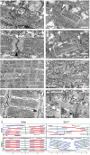
Figure 2. Sarcomeres in slou45 mutant myofibrils lack thick filaments
A-H) Electron micrographs of sarcomeres from wild-type (A,C,E,G) and slou45 mutant (B,D,F,H) myofibrils at the ages shown bottom left. A,B) Caudal myotomes of 24hpf embryos. White arrowheads (A) show immature Z-discs with poorly positioned thick and thin filaments. In slou45 mutants, bundles of thin filaments are present with putative early Z-discs (B, arrowheads). Very rarely, structures with the approximate correct dimensions of thick filaments were present (box, B). C,D) Rostral myotomes of 24hpf embryos. The wild-type has recognisable sarcomeres with almost straight Z-discs (white arrowhead, C) and more fully formed sarcoplasmic reticulum with triads (black arrowhead, C). In the slou45 mutant, I-Z-I brushes are formed (Z-discs (white arrowheads, D) flanked on either side by thin filaments), and a few putative thick filaments are evident (box, D). E-H) 48hpf wild-type muscle fibres are packed with mature myofibrils (E&G), possessing mature triads at each Z-disc (Black arrowhead, G). Mutant muscle fibres contain no thick filaments but numerous I-Z-I brushes are evident (F,H). The Z-lines are surrounded on either side by an electron-dense region (Bracket, H; F), beyond this region the thin filaments have light striations on them. Triads are present although less common (black arrowhead, H). I) A schematic summarising the principle differences between slou45 mutant and sibling sarcomere ultrastructure. At early stages of myofibrillogenesis (top) mutants have less numerous and malformed thick filaments (red) and I-Z-I brushes (green and blue) are present but less aligned than Siblings. By 48hpf, wild-type sarcomeres are fully formed whereas in mutants only mis-aligned and mis-spaced I-Z-I brushes with striated thin filaments are present (blue). T-tubules and sarcoplasmic reticulum are shown in yellow and purple. Scale bars: (A-D,G,H=500nm; E,F=2μm; H inset=100nm)
Figure 3
Figure 3. The ATPase activity of Hsp90 is abrogated by the slou45 mutation
A) Hsp90a domain structure (amino acid numbers of domain boundaries are shown below; known functions of regions indicated by black bars). The positions and consequences of mutations in three slo alleles: u45, tu44c and tm201 are shown below. The equivalent residue locations of yeast Hsp90 represented in B are shown above. B) Pymol diagram showing critical binding interactions of ADP with yeast Hsp90. Dotted blue lines are hydrogen bonds and amino acid residues (corresponding residue numbers in zebrafish in parentheses - also indicated in A) involved are in green, water molecules are cyan coloured balls and residues packed against the cα-atom of Gly83 are shown in cyan. Illustrated residues are conserved between yeast and zebrafish except Ser138/Thr149 and Ser140/His151 (illustrated in grey), which are conservative substitutions unlikely to cause major structural changes. Binding of ATP/ADP to the Hsp90 N-terminal domain involves highly conserved interactions including the carboxylate side-chain of Asp79 and main-chain carboxyl of Leu34, via a tightly bound water molecule, to the exocyclic N6 of adenine. The same Asp79 also interacts via another tightly bound water to the N1 imino nitrogen of the adenine. This same water is bound by an interaction with the side-chain hydroxyl of Thr171 and main-chain amide of Gly83. An aspartic acid residue substitution at Gly83 (mimicking the u45 mutation) would lead to steric clashes likely to disrupt critical hydrogen bonding interactions with ADP/ATP. C) ATPase activity of yeast Hsp90, human Hsp90α and their u45-mimick mutants. ATPase activity of the yeast G83D and human G91D mutants is negligible relative to corresponding wild-type.
Figure 4
Figure 4. Wildtype Hsp90a rescues thick filament formation in slou45 muscle cells whereas Hsp90au45 does not
A-D) Muscle fibres from a slou45 mutant (B,D) and sibling (A,C) injected with DNA encoding myc:WTHsp90a double stained for myc (red) and MHC (MHC;green). MHC staining in siblings decorates the cells with organised bands (C) indicative of mature A-bands. In the slou45 mutant, striations in the MHC staining are only evident in myc+ cells (D). E,F) Muscle fibres in sibling embryos injected with myc-tagged hsp90au45 DNA show organised MHC staining of myc+ cells (E). Myc+ cells in slou45 mutants injected with the same construct do not show organised myofibrils (F). Scale bars: (A,B=100μm; C-F=10μm)
Figure 5
Figure 5. Abrogation of hsp90a but not hsp90a2 function leads to upregulation of genes encoding proteins likely to be involved in sarcomere assembly
A-I) Lateral views at the stage shown bottom left showing expression of genes indicated bottom right in wild-type and mutant embryos (indicated top right). Note that although hspa8l and unc45b are upregulated in the slotu44c allele, hsp90a itself is not. J) Western blots using lysates of slou45 and slotu44c mutant and sibling embryos. More Hsp90 protein was present in slou45 mutant lysates then the corresponding siblings. In contrast, slotu44c mutant lysates contained less Hsp90 protein than siblings. K,L) Quantitative PCR (qPCR) analyses of levels of hsp90a and hsp90a2 expression in wild-type, slou45 and slotu44c mutants. Absolute expression levels of hsp90a (normalised to μg of total RNA) were significantly increased (K; p=0.002, n=5) in slou45 mutants compared to siblings; levels of expression of hsp90a in slotu44c mutants were unchanged compared to siblings (K; p=0.75, n=3); and, levels of expression of hsp90a2 were not significantly different between slou45 mutants and siblings nor slotu44c mutants and siblings (L; p=0.813 n=5; p=0.074 n=3 respectively)

Similar articles

Cited by

References

    1. Ali MM, Roe SM, Vaughan CK, Meyer P, Panaretou B, Piper PW, Prodromou C, Pearl LH. Crystal structure of an Hsp90-nucleotide-p23/Sba1 closed chaperone complex. Nature. 2006;440:1013–7. - PMC - PubMed
    1. Barral JM, Bauer CC, Ortiz I, Epstein HF. Unc-45 mutations in Caenorhabditis elegans implicate a CRO1/She4p-like domain in myosin assembly. J Cell Biol. 1998;143:1215–25. - PMC - PubMed
    1. Barral JM, Hutagalung AH, Brinker A, Hartl FU, Epstein HF. Role of the myosin assembly protein UNC-45as a molecular chaperone for myosin. Science. 2002;295:669–71. - PubMed
    1. Blagg BS, Kerr TD. Hsp90 inhibitors: small molecules that transform the Hsp90 protein folding machinery into a catalyst for protein degradation. Med Res Rev. 2006;26:310–38. - PubMed
    1. Chun M, Falkenthal S. Ifm(2)2 is a myosin heavy chain allele that disrupts myofibrillar assembly only in the indirect flight muscle of Drosophila melanogaster. J Cell Biol. 1988;107:2613–21. - PMC - PubMed

Publication types

MeSH terms