. 2008 Mar;135(6):1039-47.
doi: 10.1242/dev.017061. Epub 2008 Feb 6.

A translational block to HSPG synthesis permits BMP signaling in the early Drosophila embryo

Affiliations

A translational block to HSPG synthesis permits BMP signaling in the early Drosophila embryo

Douglas J Bornemann et al. Development. 2008 Mar.

Abstract

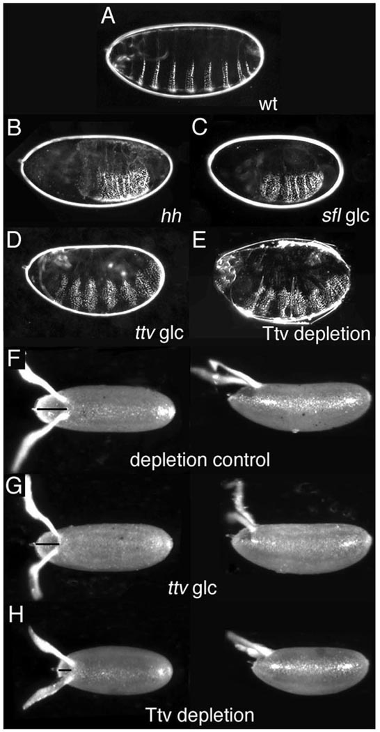
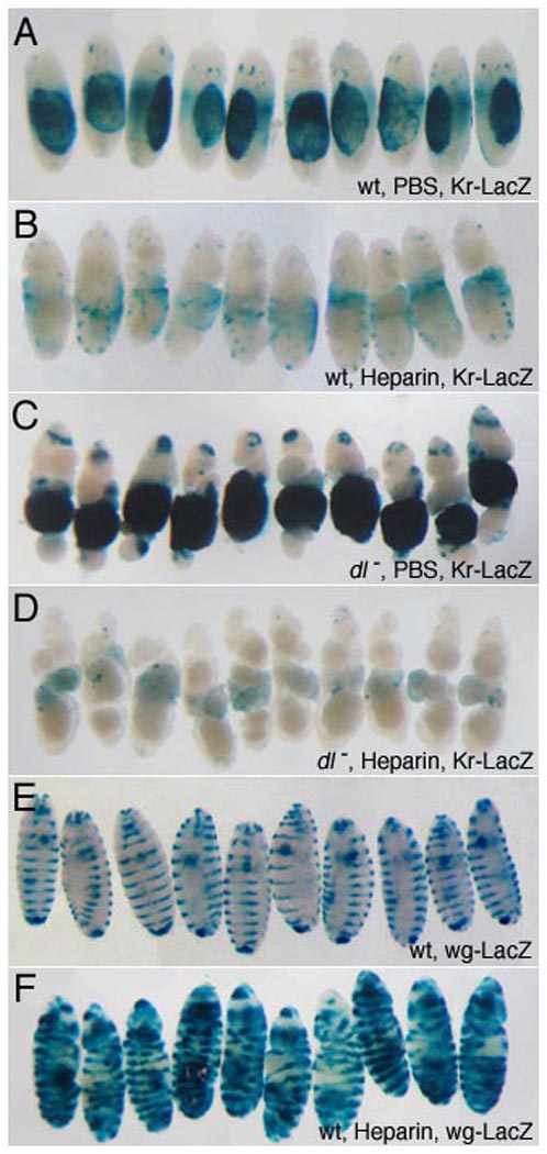
Heparan sulfate proteoglycans (HSPGs) are extracellular macromolecules found on virtually every cell type in eumetazoans. HSPGs are composed of a core protein covalently linked to glycosaminoglycan (GAG) sugar chains that bind and modulate the signaling efficiency of many ligands, including Hedgehog (Hh), Wingless (Wg) and Bone morphogenetic proteins (BMPs). Here, we show that, in Drosophila, loss of HSPGs differentially affects embryonic Hh, Wg and BMP signaling. We find that a stage-specific block to GAG synthesis prevents HSPG expression during establishment of the BMP activity gradient that is crucial for dorsal embryonic patterning. Subsequently, GAG synthesis is initiated coincident with the onset of Hh and Wg signaling which require HSPGs. This temporal regulation is achieved by the translational control of HSPG synthetic enzymes through internal ribosome entry sites (IRESs). IRES-like features are conserved in GAG enzyme transcripts from diverse organisms, suggesting that this represents a novel evolutionarily conserved mechanism for regulating GAG synthesis and modulating growth factor activity.


Figures

Figure 1
Figure 1. Embryonic Dorsal/Ventral patterning is unaffected by the absence of HSPG synthesis
(A–E) Dark field images of cuticles from (A) wild-type and (B–E) mutant larvae. (F–H) Dorsal and lateral views of eggs laid by wild-type and ttv mothers. Anterior is to the left. (A) The ventral cuticle of wild-type larvae displays a regular segmental pattern of denticle belts alternating with bands of naked cuticle. (B) Larvae mutant for hh (as well as wg) develop a continuous lawn of denticles due to loss of naked cuticle. (C) sfl germline clone (glc) larvae lacking maternal and zygotic N-deacetyl/N-sulfotransferase activity show loss of naked cuticle and fused denticles resembling the hh mutant phenotype, but no increase in denticle belt width or D/V patterning defects. (D) ttv glc larvae display a similar phenotype, with no abnormalities in D/V patterning. (E) The phenotype of a ttv mutant larva from a depleted mother that lacks somatic and germline ttv activity resembles ttv glc mutants. (F) Paired paddle-shaped dorsal appendages are located posterior to the operculum (line) at the anterior of the wild-type eggshell (G) Eggshell morphology is unaffected by loss of ttv activity in the germline (H) Eggs laid by Ttv-depleted females are shorter and have a smaller operculum, phenotypes associated with reduced Dpp signaling activity in follicle cells.
Figure 2
Figure 2. HSPG GAG chain synthesis is temporally regulated
(A) Western blots of staged embryonic extracts probed with 3G10 antisera against GAG chain stub epitopes generated by heparitinase III digestion. GAG-modified core proteins are undetectable in 0–3 hour embryo extracts but are seen in 2–4 and 4–7 hour samples. No signal was detected in the absence of heparitinase. LC denotes loading control (B) Staged embryonic extracts were probed to with anti-Dlp to detect Dlp and (C) anti-Myc to detect Dally. matTub>Gal4 was used to drive UAS-Dlp or UAS-Dally expression maternally and embryonic extracts from the indicated stages resolved on reducing gels. Dlp was detected as a sharp band at 80 kDa corresponding to full-length protein and a heterogeneous band between 50 and 60 kDa that represents a GAG-modified cleavage product. Low levels of full-length Dlp are seen at 0–3 hours, but GAG modifications are essentially undetectable until 2–4 hour and are dramatically upregulated in 4–7 hour embryos. (C) Full-length epitope-tagged Dally is visible as an ~80 kDa doublet in 0–3 and 2–4 hour extracts. Significant levels of GAG modification are first apparent in 4–7 hour extracts as decreased mobility of the full-length protein (*). The ~65 kDa band is likely to result from processing by protein convertases, similar to vertebrate glypicans.
Figure 3
Figure 3. GAG enzyme synthesis is post-transcriptionally regulated
(A) Transcripts for enzymes involved in HSPG GAG chain synthesis are maternally provided. Template RNA isolated from unfertilized eggs supports generation of RT-PCR products for all enzymes involved in HSPG GAG chain synthesis. UDP glucose dehydrogenase (Sgl) and the Glucuronic acid transporter Fringe connection (Frc) act in synthesis of GAG chain building blocks. Peptide-o-xylosyltransferase (Oxt), Galactosyltransferase I and II (GalT1, GalT2), and Glucuronyltransferases AT-1, BS1 and BS2 synthesize the tetrasaccharide linker, the polymerases Botv, Ttv and Sotv are required for chain elongation. The glypicans Dlp and Dally encode the core protein substrate for GAG chain addition and the N-deacetylase-N-sulfotransferase Sfl modifies sugar residues on the polymerised GAG chains. Dally mRNA is undetectable in eggs although an RT-PCR product can be generated using RNA from 0–24 hour embryos. To rule out amplification of a DNA template, primer pairs for all genes except galT2 (which lacks an intron) span one or more introns. (B) Western blots of staged embryonic extracts were probed to visualize expression of the indicated proteins. Loading controls are shown below each panel (i) Endogenous Sfl is absent from 0–3 hour embryos and is first detected in 2–4 hour extracts. (ii) Antisera against Ttv reveal that expression of the endogenous protein follows a similar temporal profile. (iii) In unfertilized eggs Sfl is barely detectable at 0–3 hours but is robustly expressed in 2–4 and 4–7 hour sample. (iv) Ttv expression in unfertilized eggs mirrors its expression in embryos (C, D) The ttv 5’ UTR shows IRES activity in (C) reticulocyte lysates and (D) Drosophila S2 cells. Luciferase assays were carried out on reticulocyte in vitro translation extracts or lysates from cells transfected with unmodified bicistronic vector, vector with the CV 5.1 IRES or the ttv 5’ UTR. The ratio of Renilla to Firefly luciferase was calculated for each sample and the data represented as fold amplification relative to the values for the empty pRSTF vector, set at 1. In in vitro translation extracts the ttv 5’UTR confers a 7.5 fold increase in firefly luciferase expression, comparable to the 8 fold increase generated by the CV IRES (average of two assays). In cultured cells the ttv UTR is more than three times more effective than the viral IRES and results in ~ 108 fold stimulation compared to empty vector. The graph represents the average values and standard error from 5 independent transfection assays.
Figure 4
Figure 4. Delineation of mRNA cis-elements that regulate ttv expression
(A) Western blots of staged embryonic extracts from flies transgenic for the indicated constructs were probed to visualize expression of Ttv or GFP. LC denotes loading control (i) Truncation of the 5’ UTR to eliminate upstream AUG sequences and removal of the 3’ UTR permits translation of maternally loaded Ttv-myc transcript in 0–3 hour embryos (upper band) while the endogenous protein (lower band) is not expressed at this time. (ii–v) matTub>Gal4 was used drive expression of UAS constructs in which GFP coding sequence was flanked by the indicated regions of ttv. (ii) GFP expression was detected in 0–3 hour extracts in the absence of ttv regulatory sequences as well as when only 5’ (iv) or 3’ (v) UTRs were present. (iii) In contrast, GFP expression was blocked in early embryos and initiated at ~3 hours similar to endogenous Ttv when the ORF was flanked by both 5’ and 3’ UTR sequences. (B–F) Translational regulation visualized in live animals. GFP fluorescence is absent from a female transgenic for a construct in which GFP is flanked by ttv 5’ and 3’ UTRs. (C) In contrast, ovarian expression results in bright fluorescence in a female transgenic for matTub>Gal4 and a GFP construct that lacks ttv UTRs. (D–E) Merged images showing DAPI stained nuclei and GFP fluorescence in egg chambers. (D) Egg chambers from an animal carrying matTub>Gal4 and the reporter construct with ttv 5’ and 3’ UTRs showing the absence of GFP expression and only yolk autofluorescence. (E) In contrast a reporter that lacks ttv UTRs generates high levels of GFP expression in nurse cells (nc) and oocytes (oo). Note that expression cannot be detected in follicle cells (fc). (F) Time-lapse series of a 5’ttv-UAS-GFP-3’ttv embryo shows that significant GFP fluorescence develops only after three hours post-egg laying.
Figure 5
Figure 5. Perivitelline injection of heparin inhibits Dpp signaling and disrupts embryonic dorsal/ventral patterning
Heparin/PBS solution was injected into the perivitelline space of 1–2 hour embryos to achieve the specified final concentrations (see Methods). Embryos were allowed to develop for 21 hours before fixation and staining. The dorsal-most embryonic tissue, the amnioserosa, is marked by expression of a Kr-lacZ transgene (A–D) that provides a readout of alterations in D/V patterning. Representative embryos are shown; the total sample size is indicated in parenthesis for each genotype. Injection of heparin at 1.5 µg/ml leads to reduction in Kr-LacZ expression (A, n=136, B, n=114) and morphological defects typical of loss of dorsal cell fates, such as expansion of the cephalic furrow. In dl embryos, ubiquitous dpp expression results in ventral expansion of reporter expression (C, n=40). Injected heparin (10.5 µg/ml) inhibits reporter expression in embryos lacking dl activity, demonstrating that heparin directly interferes with BMP signaling (D, n=37). In contrast, embryonic morphology along the A/P axis and segmental expression of a wg-lacZ, reporter (E), are unaffected by heparin (1.5 µg/ml), (F, n=121). This indicates that A/P patterning and Wg/Hh activity are not compromised at heparin levels that disrupt BMP signaling, providing a control for specificity. Expansion of wg-lacZ stripes laterally so that they encircle the embryo reflects cell fate changes resulting from ventralization.

References

    1. Arbeitman MN, Furlong EE, Imam F, Johnson E, Null BH, Baker BS, Krasnow MA, Scott MP, Davis RW, White KP. Gene expression during the life cycle of Drosophila melanogaster. Science. 2002;297:2270–2275. - PubMed
    1. Baeg GH, Selva EM, Goodman RM, Dasgupta R, Perrimon N. The Wingless morphogen gradient is established by the cooperative action of Frizzled and Heparan Sulfate Proteoglycan receptors. Dev Biol. 2004;276:89–100. - PubMed
    1. Binari RC, Staveley BE, Johnson WA, Godavarti R, Sasisekharan R, Manoukian AS. Genetic evidence that heparin-like glycosaminoglycans are involved in wingless signaling. Development. 1997;124:2623–2632. - PubMed
    1. Bishop JR, Schuksz M, Esko JD. Heparan sulphate proteoglycans fine-tune mammalian physiology. Nature. 2007;446:1030–1037. - PubMed
    1. Bornemann DJ, Duncan JE, Staatz W, Selleck S, Warrior R. Abrogation of heparan sulfate synthesis in Drosophila disrupts the Wingless, Hedgehog and Decapentaplegic signaling pathways. Development. 2004;131:1927–1938. - PubMed

Publication types

MeSH terms