

Diffusion coefficient of fluorescent phosphatidylinositol 4,5-bisphosphate in the plasma membrane of cells
- PMID: 18256277
- PMCID: PMC2291420
- DOI: 10.1091/mbc.e07-12-1208
Diffusion coefficient of fluorescent phosphatidylinositol 4,5-bisphosphate in the plasma membrane of cells
Abstract
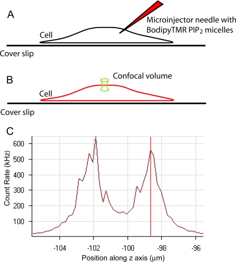
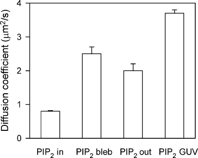
Phosphatidylinositol 4,5-bisphosphate (PIP(2)) controls a surprisingly large number of processes in cells. Thus, many investigators have suggested that there might be different pools of PIP(2) on the inner leaflet of the plasma membrane. If a significant fraction of PIP(2) is bound electrostatically to unstructured clusters of basic residues on membrane proteins, the PIP(2) diffusion constant, D, should be reduced. We microinjected micelles of Bodipy TMR-PIP(2) into cells, and we measured D on the inner leaflet of fibroblasts and epithelial cells by using fluorescence correlation spectroscopy. The average +/- SD value from all cell types was D = 0.8 +/- 0.2 microm(2)/s (n = 218; 25 degrees C). This is threefold lower than the D in blebs formed on Rat1 cells, D = 2.5 +/- 0.8 microm(2)/s (n = 26). It is also significantly lower than the D in the outer leaflet or in giant unilamellar vesicles and the diffusion coefficient for other lipids on the inner leaflet of these cell membranes. The simplest interpretation is that approximately two thirds of the PIP(2) on inner leaflet of these plasma membranes is bound reversibly.
Figures

Comment in
- Mol Biol Cell. 19:1281.
References
-
- Almeida P. F., Vaz W.L.C. Lateral diffusion in membranes. In: Lipowsky R., Sackmann E., editors. Handbook of biological physics. Amsterdam, The Netherlands: Elsevier Science; 1995. pp. 305–357.
-
- Anderson R. A., Marchesi V. T. Regulation of the association of membrane skeletal protein 4.1 with glycophorin by a polyphosphoinositide. Nature. 1985;318:295–298. - PubMed
Publication types
MeSH terms
Substances
Grants and funding
LinkOut - more resources
Full Text Sources
Other Literature Sources
