Comparative Study
. 2008 Apr;19(4):1540-7.
doi: 10.1091/mbc.e07-09-0942. Epub 2008 Feb 6.

Identification of ciliary localization sequences within the third intracellular loop of G protein-coupled receptors

Affiliations
Comparative Study

Identification of ciliary localization sequences within the third intracellular loop of G protein-coupled receptors

Nicolas F Berbari et al. Mol Biol Cell. 2008 Apr.

Abstract

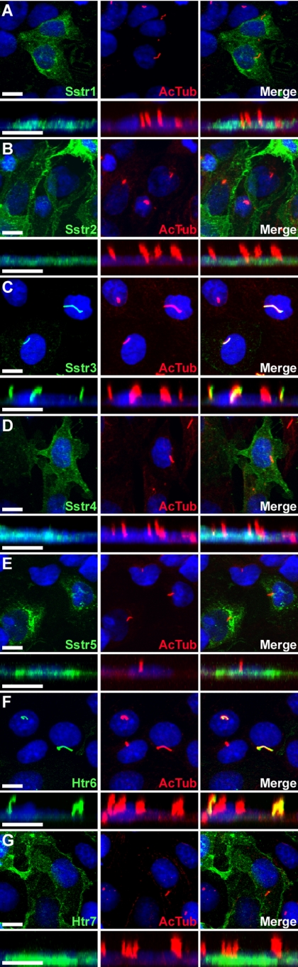
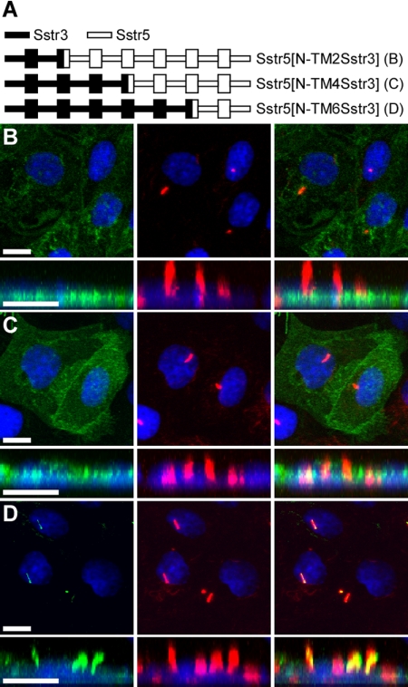
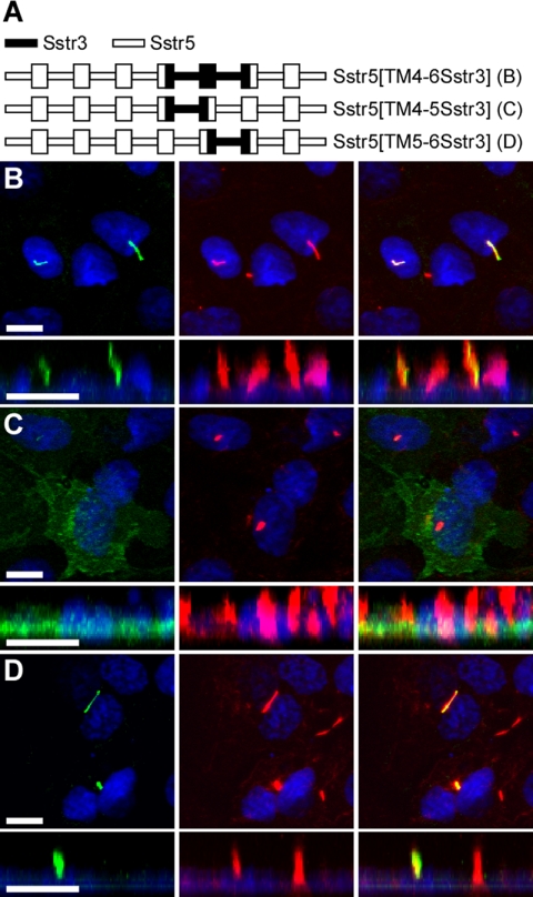
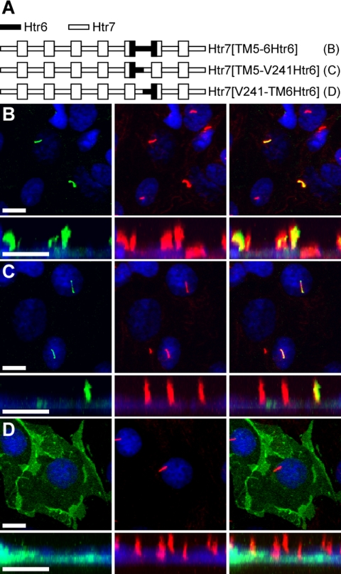
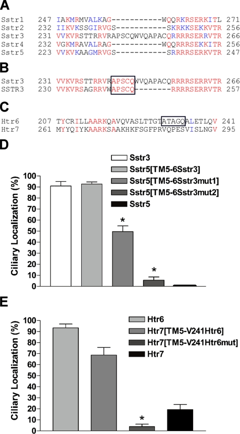
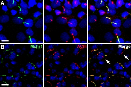
Primary cilia are sensory organelles present on most mammalian cells. The functions of cilia are defined by the signaling proteins localized to the ciliary membrane. Certain G protein-coupled receptors (GPCRs), including somatostatin receptor 3 (Sstr3) and serotonin receptor 6 (Htr6), localize to cilia. As Sstr3 and Htr6 are the only somatostatin and serotonin receptor subtypes that localize to cilia, we hypothesized they contain ciliary localization sequences. To test this hypothesis we expressed chimeric receptors containing fragments of Sstr3 and Htr6 in the nonciliary receptors Sstr5 and Htr7, respectively, in ciliated cells. We found the third intracellular loop of Sstr3 or Htr6 is sufficient for ciliary localization. Comparison of these loops revealed a loose consensus sequence. To determine whether this consensus sequence predicts ciliary localization of other GPCRs, we compared it with the third intracellular loop of all human GPCRs. We identified the consensus sequence in melanin-concentrating hormone receptor 1 (Mchr1) and confirmed Mchr1 localizes to primary cilia in vitro and in vivo. Thus, we have identified a putative GPCR ciliary localization sequence and used this sequence to identify a novel ciliary GPCR. As Mchr1 mediates feeding behavior and metabolism, our results implicate ciliary signaling in the regulation of body weight.


Figures

Figure 1.
Figure 1.
Somatostatin receptor subtype 3 and serotonin receptor subtype 6 selectively localize to cilia when heterologously expressed in IMCD cells. (A–G) Representative confocal microscopy images of transiently transfected IMCD cells expressing somatostatin receptors 1 through 5 (Sstr1–5) and serotonin receptors 6 (Htr6) and 7 (Htr7) fused at the C-terminus to EGFP. Left, EGFP fluorescence (green) shows expression of the receptors; middle, acetylated α-tubulin (red) marks the cilia; right, merged images. Below each panel is a confocal image in the xz plane to show cilia projecting vertically from the apical surface of cells. Note that only Sstr3-EGFP (C) and Htr6-EGFP (F) show localization that overlaps with acetylated α-tubulin ciliary labeling. Sstr2 corresponds to Sstr2a. Nuclei are labeled with the DNA stain DRAQ5 (blue). All scale bars, 10 μm.
Figure 2.
Figure 2.
Sequences between the fourth and sixth transmembrane domains of Sstr3 are important for ciliary localization. (A) Schematic of chimeric receptors containing portions of Sstr3 (indicated by black lines) and Sstr5 (indicated by white lines) fused at the C-terminus to EGFP. Transmembrane domains (TM) are depicted as boxes. (B–D) Representative images of transiently transfected IMCD cells expressing the indicated chimeric receptors. Left, EGFP fluorescence (green); middle, acetylated α-tubulin (red); right, merged images. Chimeric receptors Sstr5[N-TM2Sstr3] (B) and Sstr5[N-TM4Sstr3] (C) do not localize to cilia. Chimeric receptor Sstr5[N-TM6Sstr3] does localize to cilia, suggesting that ciliary localization of Sstr3 is mediated by sequences between TM4 and TM6. Nuclei are labeled with DRAQ5 (blue). All scale bars, 10 μm.
Figure 3.
Figure 3.
The third intracellular (i3) loop of Sstr3 is sufficient to localize Sstr5 to cilia. (A) Schematic of chimeric receptors containing portions of Sstr3 (black lines) and Sstr5 (white lines) fused at the C-terminus to EGFP. TM domains are depicted as boxes. (B–D) Representative images of transiently transfected IMCD cells expressing the indicated chimeric receptors. Left, EGFP fluorescence (green); middle, acetylated α-tubulin (red); right, merged images. Chimeric receptors Sstr5[TM4-6Sstr3] (B) and Sstr5[TM5-6Sstr3] (D) localize to cilia, whereas chimeric receptor Sstr5[TM4-5Sstr3] (C) does not localize to cilia, suggesting that the i3 loop of Sstr3 contains ciliary localization sequences. Nuclei are labeled with DRAQ5 (blue). All scale bars, 10 μm.
Figure 4.
Figure 4.
The amino portion of the i3 loop of Htr6 is sufficient to localize Htr7 to cilia. (A) Schematic of chimeric receptors containing portions of Htr6 (black lines) and Htr7 (white lines) fused at the C-terminus to EGFP. TM domains are depicted as boxes. (B–D) Representative images of transiently transfected IMCD cells expressing the indicated chimeric receptors. Left, EGFP fluorescence (green); middle, acetylated α-tubulin (red); right, merged images. Chimeric receptors Htr7[TM5-6Htr6] (B) and Htr7[TM5-V241Htr6] (C) selectively localize to cilia, whereas chimeric receptor Htr7 [V241-TM6Htr6] (D) does not, suggesting that the N-portion of the i3 loop of Htr6 contains ciliary localization sequences. Nuclei are labeled with DRAQ5 (blue). All scale bars, 10 μm.
Figure 5.
Figure 5.
Comparative genomics and mutational analysis identifies unique residues in Sstr3 and Htr6 that are important for ciliary localization of GPCRs. (A) Alignment of the predicted i3 loop sequences of the mouse somatostatin receptors reveals an insertion of 11 unique residues in Sstr3. The corresponding positions of the residues are indicated. Red signifies identical residues and blue signifies conserved residues. (B) Alignment of the predicted i3 loop sequence of somatostatin receptor subtype 3 from mouse (Sstr3) and human (SSTR3). Five of the unique 11 residues (APSCQ; boxed) are completely conserved between mouse and human. (C) Alignment of the predicted i3 loop sequences of mouse serotonin receptors 6 and 7 shows the presence of a unique sequence in Htr6 (ATAGQ; boxed) with modest similarity to the Sstr3 sequence. (D) Percentage of transiently transfected IMCD cells that show ciliary localization when expressing Sstr3, Sstr5, Sstr5[TM5-6Sstr3], Sstr5[TM5-6Sstr3mut1], or Sstr5[TM5-6Sstr3mut2]. Sstr3 localizes to cilia in ∼91% of transfected cells. Sstr5 never localizes to cilia. Chimeric receptor Sstr5[TM5-6Sstr3] localizes to cilia in ∼93% of transfected cells. Chimeric receptor Sstr5[TM5-6Sstr3mut1], in which the A and Q in the conserved consensus sequence have been mutated to F, localizes to cilia in ∼50% of transfected cells. Chimeric receptor Sstr5[TM5–6Sstr3mut2], in which the A and Q in the second consensus sequence have also been mutated to F, localizes to cilia in ∼6% of transfected cells. Values are expressed as mean ± SEM *Significantly different from Sstr3 and Sstr5[TM5–6Sstr3] percentages. (E) Percentage of transiently transfected IMCD cells that show ciliary localization when expressing Htr6, Htr7, Htr7[TM5-V241Htr6], or Htr7[TM5-V241Htr6mut]. Htr6 localizes to cilia in ∼93% of transfected cells. Htr7 localizes to cilia in ∼20% of transfected cells. Chimeric receptor Htr7[TM5-V241Htr6] localizes to cilia in ∼70% of transfected cells. Chimeric receptor Htr7[TM5-V241Htr6mut], in which the A and Q have been mutated to F, localizes to cilia in ∼4% of transfected cells. Values are expressed as mean ± SEM. *Significantly different from Htr6 and Htr7[TM5-V241Htr6] percentages.
Figure 6.
Figure 6.
Melanin-concentrating hormone receptor 1 (Mchr1) localizes to cilia in vitro and in vivo. (A) Representative image of transiently transfected IMCD cells expressing Mchr1 fused at the C-terminus to EGFP. Left, EGFP fluorescence (green); middle, acetylated α-tubulin (red); right, merged image. (B) Representative image of the nucleus accumbens from an adult mouse colabeled with antibodies to Mchr1 (green; left), ACIII (red; middle), and merged (right). The majority of cilia are positive for both Mchr1 and ACIII. Cilia that are positive for ACIII but negative for Mchr1 are indicated with arrows. Nuclei are labeled with DRAQ5 (blue). All scale bars, 10 μm.

References

    1. Badano J. L., Mitsuma N., Beales P. L., Katsanis N. The ciliopathies: an emerging class of human genetic disorders. Annu. Rev. Genomics Hum. Genet. 2006;7:125–148. - PubMed
    1. Berbari N. F., Bishop G. A., Askwith C. C., Lewis J. S., Mykytyn K. Hippocampal neurons possess primary cilia in culture. J. Neurosci. Res. 2007;85:1095–1100. - PubMed
    1. Bisgrove B. W., Yost H. J. The roles of cilia in developmental disorders and disease. Development. 2006;133:4131–4143. - PubMed
    1. Bishop G. A., Berbari N. F., Lewis J. S., Mykytyn K. Type III adenylyl cyclase localizes to primary cilia throughout the adult mouse brain. J. Comp. Neurol. 2007;505:562–571. - PubMed
    1. Bloodgood R. A. Protein targeting to flagella of trypanosomatid protozoa. Cell Biol. Int. 2000;24:857–862. - PubMed

Publication types

MeSH terms