. 2008 Apr;19(4):1727-38.
doi: 10.1091/mbc.e07-07-0718. Epub 2008 Feb 6.

Schizosaccharomyces pombe Pxl1 is a paxillin homologue that modulates Rho1 activity and participates in cytokinesis

Affiliations

Schizosaccharomyces pombe Pxl1 is a paxillin homologue that modulates Rho1 activity and participates in cytokinesis

Mario Pinar et al. Mol Biol Cell. 2008 Apr.

Abstract

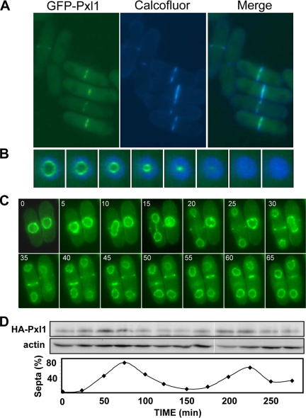
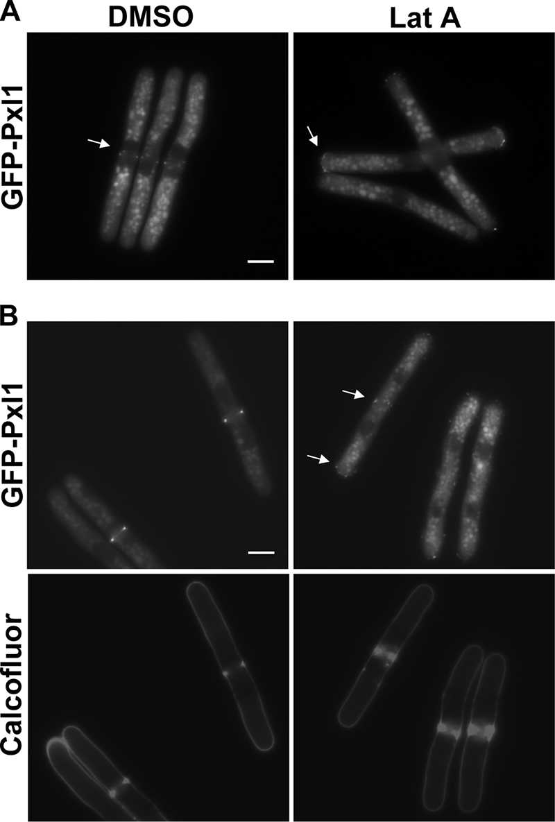
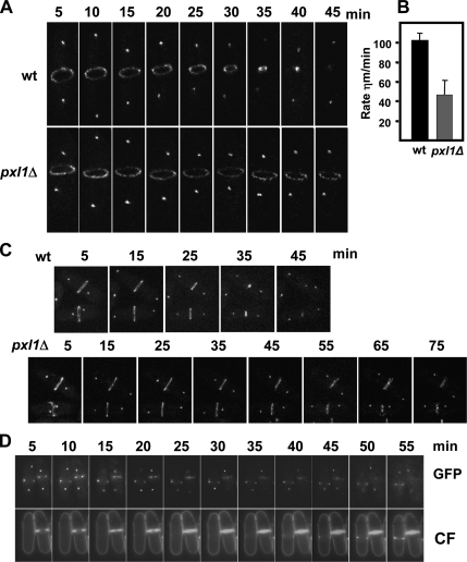
Schizosaccharomyces pombe Rho GTPases regulate actin cytoskeleton organization and cell integrity. We studied the fission yeast gene SPBC4F6.12 based on its ability to suppress the thermosensitivity of cdc42-1625 mutant strain. This gene, named pxl1(+), encodes a protein with three LIM domains that is similar to paxillin. Pxl1 does not interact with Cdc42 but it interacts with Rho1, and it negatively regulates this GTPase. Fission yeast Pxl1 forms a contractile ring in the cell division region and deletion of pxl1(+) causes a delay in cell-cell separation, suggesting that it has a function in cytokinesis. Pxl1 N-terminal region is required and sufficient for its localization to the medial ring, whereas the LIM domains are necessary for its function. Pxl1 localization requires actin polymerization and the actomyosin ring, but it is independent of the septation initiation network (SIN) function. Moreover, Pxl1 colocalizes and interacts with Myo2, and Cdc15, suggesting that it is part of the actomyosin ring. Here, we show that in cells lacking Pxl1, the myosin ring is not correctly assembled and that actomyosin ring contraction is delayed. Together, these data suggest that Pxl1 modulates Rho1 GTPase signaling and plays a role in the formation and contraction of the actomyosin ring during cytokinesis.


Figures

Figure 1.
Figure 1.
S. pombe pxl1+ overexpression suppresses the thermosensitive growth of cdc42-1625. (A) cdc42-1625 cells transformed with pREP81X plasmid containing different genes: cdc42+; gef1+; scd1+; shk1+; shk2+; and pxl1+. Cells were grown at 25°C and at 36°C in media with or without thiamine to repress or induce, respectively, the nmt1+ promoter. (B) Differential interphase contrast images of cdc42-1625 cells transformed with empty pREP81X or pREP81X-pxl1+ and grown 16 h without thiamine at 25 or 36°C.
Figure 2.
Figure 2.
Pxl1 is a negative regulator of Rho1. (A) Pxl1 interacts with Rho1. Cells expressing GFP-pxl1+ and HA-rho1+ from endogenous promoters were lysated, and immunoprecipitation was performed with anti-GFP antibody and protein A-Sepharose beads. Immunoprecipitates were analyzed by Western blot by using anti-HA (bottom) antibody. The lysates were analyzed by Western blot with anti-GFP (top) or anti-HA (middle) antibodies. (B) Differential interphase contrast and fluorescence micrographs of calcofluor-stained wild-type cells (top) and pxl1Δ cells (bottom) grown at 25°C. The arrow points to a misplaced septum. The bar corresponds to 5 μm. (C) Pxl1 modulates the amount of GTP-bound Rho1. Wild-type, rga5Δ, and pxl1Δ cells expressing HA-rho1+ from its promoter were precipitated with GST-RBD and blotted with anti-HA antibodies (top). Total HA-Rho1 in cell lysates was visualized by Western blot (middle). Data were quantified and presented as percentage relative to the wild-type extracts run in the same experiment (bottom). (D) Percentages of septating and multiseptated cells in cultures of wild-type and pxl1Δ cells transformed with pREP41 plasmids carrying the indicated genes. Cells were grown 16 h without thiamine at 28°C to allow overexpression. Percentages of septating wild-type and pxl1Δ cells carrying simultaneously the rga5Δ mutation are also included. (E) The lack of Pxl1 rescues the growth at 37°C of cells carrying the rgf3+ thermosensitive allele ehs2-1. Cells were grown in rich medium for 10 h. Initial OD600 was 0.1. (F) Same cells as in E were grown in rich medium at 32°C supplemented with 50 mM NaF for 2 d. Initial OD600 was 2 and 1:4 dilutions were successively made.
Figure 3.
Figure 3.
Pxl1 localization and levels during cell cycle. (A) Fluorescence microscopy of log phase GFP-pxl1+ cells stained with calcofluor. (B) Three-dimensional projection of a time-lapse of GFP-pxl1+ cells stained with calcofluor. Pictures were taken at 5-min intervals. (C) Time-lapse fluorescence microscopy of cells expressing GFP-pxl1+ and cut11+-GFP grown to early log phase at 25°C. Pictures were taken at 5-min intervals. (D) cdc25-22 cells carrying endogenously expressed HA-pxl1+ were synchronized at 36°C for 4 h, and then they were transferred to 25°C, and samples for protein analysis were taken every 25 min. Proteins from cell extracts were analyzed by Western blot with monoclonal anti-HA and anti-actin antibodies. The percentage of septating cells in each sample is presented as an indicator of cell cycle progression.
Figure 4.
Figure 4.
Pxl1 localization depends on the N-terminal region but Pxl1 function requires the LIM domains. (A) Scheme and protein levels of GFP-Pxl1 truncations expressed under the control of pxl1+ promoter in pxl1Δ cells. The proteins were detected by Western-Blot using anti-GFP antibody and 7.5% SDS-PAGE. Molecular weight standards (right), and molecular weight of the different GFP-Pxl1 truncations (right) are indicated (B) Percentages of cells containing a single septum and multiple septa calculated from 500 pxl1Δ cells expressing different GFP-Pxl1 fragments. (C) Calcofluor and GFP fluorescence micrographs of pxl1Δ cells carrying different pxl1 fragments.
Figure 5.
Figure 5.
Pxl1 localization depends on actin polymerization. cdc25-22 GFP-pxl1+ cells grown at 25°C to mid-log phase, and then they were shifted to 36°C for 4 h and shifted back to 25°C. At time 0 (A) and 40 min (B), 1% DMSO or 50 μM Lat A was added. Cells were stained with calcofluor, and then they were analyzed by fluorescence microscopy 20 min after the addition of Lat A. The arrows point the GFP-Pxl1. Bar, 5 μm.
Figure 6.
Figure 6.
Pxl1 colocalizes with actomyosin ring proteins and physically interacts with Rlc1 and Cdc15. Cells containing cherry RFP-Pxl1 and GFP-Myo2 Sad1-GFP (A), Cdc12-3YFP (B), or GFP-Cdc15 (C) were grown to log phase, and then they were examined by fluorescence microscopy. A separated cell (inset) is shown to see the localization at early stages of cytokinesis. (D) Interaction of Rlc1-GFP and GST-Pxl1. Extracts of cells expressing rlc1+-GFP and GST-pxl1+ at physiological levels were immunoprecipitated with GS-beads and probed with anti-GFP antibodies. Extracts were assayed for the level of GST-Pxl1 and Rlc1-GFP by Western blot. (E) Interaction of GFP-cdc15 and HA-Pxl1. Extracts of cells carrying GFP-cdc15+ and HA-pxl1+ expressed at endogenous levels were immunoprecipitated with anti-HA antibodies and probed with anti-GFP antibodies. Extracts were assayed for levels of HA-pxl1 and GFP-Cdc15 by Western blot.
Figure 7.
Figure 7.
Pxl1 is required for proper myosin II localization to the actomyosin ring. (A) Fluorescence microscopy of wild-type and pxl1Δ cells carrying GFP-myo2+ sad1+-GFP, rlc1+-GFP, and cdc4+-GFP endogenously expressed. (B) Confocal fluorescence images of GFP-Myo2 and Sad1-GFP in pxl1Δ cells. (C) Fluorescence microscopy of a calcofluor-stained pxl1Δ cell expressing rlc1-GFP to see the septum formation while one of the myosin rings was not contracted. (D) Localization of Myo3-GFP in wild-type and pxl1Δ cells. Arrows point to irregular rings.
Figure 8.
Figure 8.
Ring constriction during cytokinesis is delayed in pxl1Δ cells. Analysis of Myo2 ring contraction in wild-type and pxl1Δ cells grown at 25°C. (A) Time-lapse confocal fluorescence microscopy of GFP-Myo2 Sad1-GFP in wild-type and pxl1Δ cells. (B) Rate of ring constriction at 25°C of wild type (n = 10) and pxl1Δ cells (n = 20) calculated from the time-lapse confocal images in three independent experiments. (C and D) Time-lapse fluorescence images of different GFP-Myo2 Sad1-GFP in wild-type and pxl1Δ cells during cytokinesis. In D, cells were stained with calcofluor.

Comment in

  • Mol Biol Cell. 19:1281.

References

    1. Alonso-Nunez M. L., An H., Martin-Cuadrado A. B., Mehta S., Petit C., Sipiczki M., del Rey F., Gould K. L., de Aldana C. R. Ace2p controls the expression of genes required for cell separation in Schizosaccharomyces pombe. Mol. Biol. Cell. 2005;16:2003–2017. - PMC - PubMed
    1. An H., Morrell J. L., Jennings J. L., Link A. J., Gould K. L. Requirements of fission yeast septins for complex formation, localization and function. Mol. Biol. Cell. 2004;15:5551–5564. - PMC - PubMed
    1. Arellano M., Coll P. M., Pérez P. Rho GTPases in the control of cell morphology, cell polarity, and actin localization in fission yeast. Microsc. Res. Tech. 1999a;47:51–60. - PubMed
    1. Arellano M., Duran A., Perez P. Rho 1 GTPase activates the (1–3)beta-D-glucan synthase and is involved in Schizosaccharomyces pombe morphogenesis. EMBO J. 1996;15:4584–4591. - PMC - PubMed
    1. Arellano M., Duran A., Perez P. Localization of the Schizosaccharomyces pombe Rho1 GTPase and its involvement in the organization of the actin cytoskeleton. J. Cell Sci. 1997;110:2547–2555. - PubMed

Publication types

MeSH terms