. 2008 Apr;19(4):1706-16.
doi: 10.1091/mbc.e07-07-0637. Epub 2008 Feb 6.

A novel role for PA28gamma-proteasome in nuclear speckle organization and SR protein trafficking

Affiliations

A novel role for PA28gamma-proteasome in nuclear speckle organization and SR protein trafficking

Véronique Baldin et al. Mol Biol Cell. 2008 Apr.

Abstract

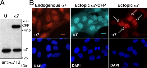
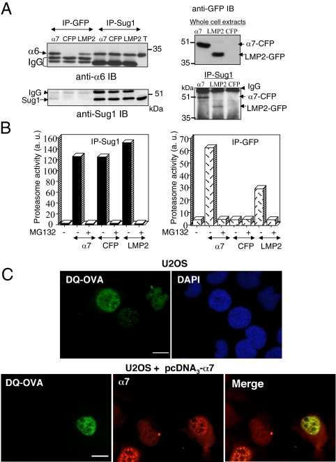
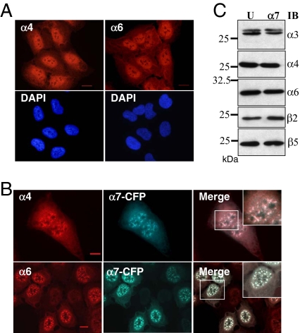
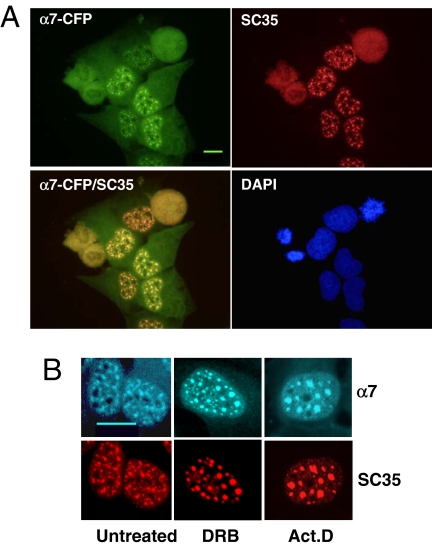
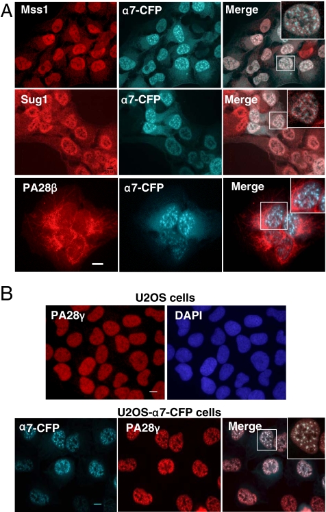
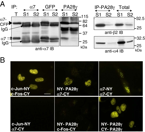
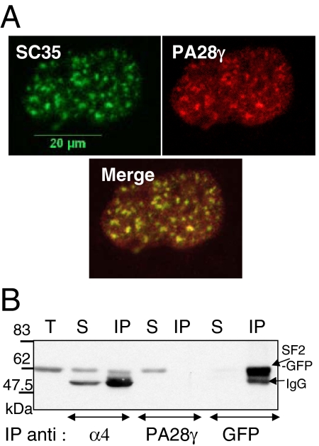
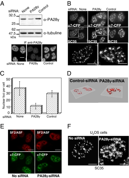
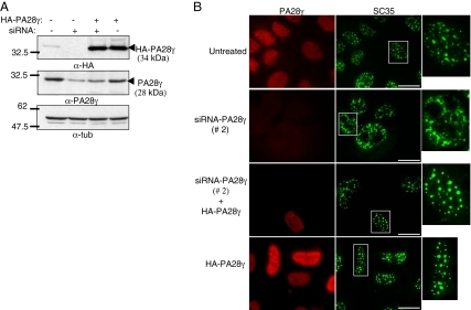
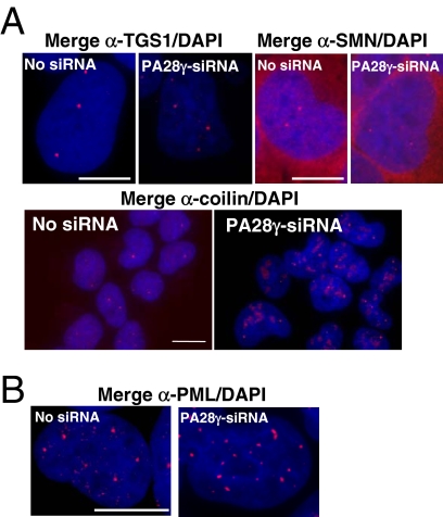
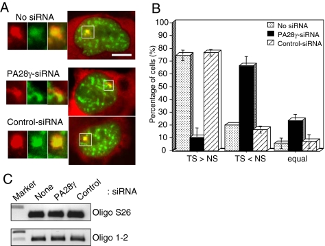
In eukaryotic cells, proteasomes play an essential role in intracellular proteolysis and are involved in the control of most biological processes through regulated degradation of key proteins. Analysis of 20S proteasome localization in human cell lines, using ectopic expression of its CFP-tagged alpha7 subunit, revealed the presence in nuclear foci of a specific and proteolytically active complex made by association of the 20S proteasome with its PA28gamma regulator. Identification of these foci as the nuclear speckles (NS), which are dynamic subnuclear structures enriched in splicing factors (including the SR protein family), prompted us to analyze the role(s) of proteasome-PA28gamma complexes in the NS. Here, we show that knockdown of these complexes by small interfering RNAs directed against PA28gamma strongly impacts the organization of the NS. Further analysis of PA28gamma-depleted cells demonstrated an alteration of intranuclear trafficking of SR proteins. Thus, our data identify proteasome-PA28gamma complexes as a novel regulator of NS organization and function, acting most likely through selective proteolysis. These results constitute the first demonstration of a role of a specific proteasome complex in a defined subnuclear compartment and suggest that proteolysis plays important functions in the precise control of splicing factors trafficking within the nucleus.


Figures

Figure 1.
Figure 1.
Ectopic α7-CFP and α7 accumulate into nuclear foci. (A) Expression of the α7-CFP protein in cells. Total cellular extracts (30 μg) of human osteosarcoma U2OS-tTA (U) or U2OS-tTA cells stably transfected with vectors conditionally expressing α7-CFP (α7) cultured in the absence of tetracycline for 48 h were separated by electrophoresis and analyzed by immunoblotting using anti-α7 antibodies. (B) The cellular distribution of α7 in U2OS-tTA cells either untransfected (endogenous α7), stably transfected with pTRE2-α7-CFP (ectopic α7-CFP), or transiently transfected with pcDNA3-α7 (ectopic α7) was analyzed by indirect immunofluorescence using anti-α7 antibodies (red) or by CFP fluorescence (cyan) on fixed cells. Arrows indicate transiently transfected cells. Observation with a 63× objective. Bar, 10 μm.
Figure 2.
Figure 2.
α7-CFP is incorporated into active proteasome complexes that accumulate in nuclear foci. (A) Total cell lysate from human osteosarcoma U2OS-tTA cells stably transfected with vectors conditionally expressing α7-CFP (α7) or CFP (CFP) cultured in the absence of tetracycline for 48 h and from MelJuso cells stably expressing LMP2-GFP (LMP2) were subjected to immunoprecipitation (IP) with anti-GFP or anti-Sug1 antibodies. Immunoprecipitated proteins as well as 30 μg of U2OS-tTA total extract (T) were separated by electrophoresis and analyzed by immunoblotting using the indicated antibodies. (B) Proteins immunoprecipitated with either anti-Sug1 or anti-GFP antibodies from total cell extracts were tested for proteasomal activity using the peptide suc-LLVY-AMC as a substrate, in the presence (+) or not (−) of 50 μM MG132 (first bar: no IP in the assay). (C) Proteasomes are active in nuclear foci. U2OS-tTA cells (top panel) or U2OS-tTA cells transiently expressing α7 (bottom panel) were microinjected (24 h after transfection) with the fluorogenic substrate protein DQ-OVA (0.5 mg/ml). The substrate was injected into the nuclei. Cells were incubated 10 min at 37°C and then fixed, and the fluorescent DQ-OVA degradation products were visualized by the appearance of green fluorescence. α7 was detected by indirect immunofluorescence with anti-α7 (red; 63× objective). Bar, 10 μm. U2OS-tTA-α7-CFP cells were not used in this experiment because of the overlap of the fluorescence spectrums of CFP and of DQ-OVA degradation products.
Figure 3.
Figure 3.
Ectopic expression of α7-CFP promotes recruitment of whole 20S proteasomes into the nuclear foci. Parental U2OS-tTA cells (A) and U2OS-tTA-α7-CFP cells induced for 48 h for α7-CFP expression (B) were subjected to indirect immunofluorescence using antibodies directed against the α4 and α6 subunits of the 20S proteasome (red). α7-CFP fusion protein was detected by CFP fluorescence. Wide-field overlay images and enlargements of marked area are shown. All observations were done with a 63× objective. Bar, 10 μm. (C) Expression of α7-CFP does not alter expression level of other 20S proteasome subunits. Thirty micrograms of total protein extract from U2OS-tTA (U) and induced U2OS-tTA-α7-CFP (α7) cells were separated by electrophoresis and analyzed by immunoblot (IB) using the indicated antibodies.
Figure 4.
Figure 4.
The nuclear foci enriched in 20S proteasome-α7-CFP complexes correspond to the NS. (A) U2OS-tTA-α7-CFP were fixed 48 h after α7-CFP induction, permeabilized, and stained with a mAb directed against the phosphorylated NS protein SC35 (red) and with DAPI dye (blue). α7-CFP fluorescence is in green for easier codetection (α7-CFP/SC35 image). Wide-field of individual detection and overlay images are presented (63× objective). Bar, 10 μm. (B) Forty-eight-hour–induced U2OS-tTA-α7-CFP cells, untreated or treated with DRB (100 μM) or actinomycin D (10 μg/ml) for 2 h before fixation, were stained with anti-SC35 (red); α7-CFP expression was directly visualized by CFP fluorescence (cyan; 100× objective). Bar, 10 μm.
Figure 5.
Figure 5.
PA28γ, but not other 20S proteasome regulatory complexes, is recruited together with 20S proteasome into the nuclear foci. (A) U2OS-tTA-α7-CFP cells induced for 48 h were analyzed by indirect immunofluorescence using antibodies directed against the Mss1/Rpt1 and Sug1/Rpt6 ATPase subunits of the 19S complex (red) or PA28β. α7-CFP fusion protein was detected by CFP fluorescence. (B) Subcellular localization of PA28γ (indirect immunofluorescence, red) in parental U2OS-tTA cells and in induced U2OS-tTA-α7-CFP cells. α7-CFP was detected by direct fluorescence. Wide field of individual areas of detection, overlay images, and enlargements of marked area are presented. All observations were done with a 63× objective. Bar, 10 μm.
Figure 6.
Figure 6.
α7 and PA28γ are physically associated in total cell extracts, in a nuclear foci-enriched fraction and in cellulo. (A) After lysis of induced U2OS-tTA-α7-CFP cells, the cell extract was clarified by centrifugation, and the supernatant was collected (S1). The insoluble material was then resuspended in lysis buffer containing 0.3% Triton X-100 (15 min at 4°C) and clarified by centrifugation, yielding the S2 supernatant. Proteins immunoprecipitated from the S1 and S2 supernatants, using the indicated antibodies, were then separated by SDS-PAGE and analyzed by immunoblotting using antibodies directed against the proteasome subunits α7 (left panel), β2 (right top panel) or α4 (right bottom panel). T: 30 μg of total proteins. (B) The interaction between α7-CFP and PA28γ was visualized in cellulo by BiFC. Proteins indicated in each panel were coexpressed in HeLa cells. Twenty-four hours after transfection of expression vectors, cells were incubated at 32°C for a further 24 h to promote chromophore maturation and then fixed to allow YFP fluorescence monitoring. c-Fos (118–210) and c-Jun (257–318) constructs (Hu et al., 2002) were used as positive (Jun-NY/Fos-CY) or negative (Jun-NY/α7-CY and NY-PA28γ/Fos-CY) BiFC controls (63× objective). Bar, 10 μm.
Figure 7.
Figure 7.
Presence of PA28γ in the NS without α7-CFP expression and association of 20S proteasome with SF2/ASF protein. (A) Parental U2OS-tTA cells were permeabilized and fixed simultaneously to remove most of soluble PA28γ complexes. Cells were then stained with anti-SC35 (green) and anti-PA28γ (red), and an overlay image is shown (100× objective). (B) U2OS-tTA cells, cotransfected with pcDNA3-α7 and pEGFP-SF2/ASF vectors, were lysed 24 h after transfection in lysis buffer containing 0.3% Triton X-100. Two hundred micrograms of total cell extract were subjected to immunoprecipitation using either anti-α4, anti-PA28γ or anti-GFP antibodies. Immunoprecipitated proteins were then separated by SDS-PAGE (10%) and visualized by immunoblotting using anti-GFP antibody. T, 30 μg of total cells extract; S, supernatant of the IP (1/10); IP, immunoprecipitation.
Figure 8.
Figure 8.
Proteasome-PA28γ complexes are required for the integrity of the NS. (A) U2OS-tTA-α7-CFP cells were induced for α7-CFP expression and concomitantly transfected with control-siRNA or PA28γ-siRNA (siRNA 1) duplexes. Cells were recovered 48 h after transfection, and the expression level of PA28γ was analyzed either by immunoblotting or by indirect immunofluorescence using an anti-PA28γ antibody. Bar, 10 μm. (B) NS were visualized by the fluorescence of α7-CFP and by indirect immunofluorescence using anti-SC35 antibodies in induced U2OS-tTA-α7-CFP cells untransfected or transfected with PA28γ- or control-siRNA duplexes. Enlargements of the marked areas are presented. Bar, 10 μm. (C) Quantification of the number of α7-CFP–labeled NS in cells treated with PA28γ- or control-siRNA duplexes. Quantification was performed visually, by counting on the pictures the number of compact fluorescent foci in each cell. The values correspond to the means of five independent experiments (n = 100 cells, ±SD). (D) 3D reconstruction. SC35 localization was detected by indirect immunofluorescence in induced U2OS-tTA-α7-CFP cells treated with control- or PA28γ (1)-siRNA. Fixed cells were observed with a Leica DMRA microscope equipped with a 63× PL APO (NA = 1.32) oil immersion objective and a N2.1 (Leica) filter set. Stacks of images were acquired using a piezo stepper (Physik Instruments, Waldbronn, Germany), Metamorph 7.1 (Molecular Devices, Menlo Park, CA) and a Micromax 1300YHS CCD camera (Princeton Research Instruments, Princeton, NJ). Stacks were further deconvolved using a MLE algorithm and the Huygens 2.9 software (Scientific Volume Imaging, Hilversrum, The Netherlands) and analyzed in 3D using Imaris 5.3 (Bitplane, Zurich, Switzerland). (E) NS were visualized by the fluorescence of α7-CFP and by indirect immunofluorescence using anti-SF2/ASF antibodies in induced U2OS-tTA-α7-CFP cells untransfected or transfected with PA28γ- or control-siRNA duplexes. Bar, 10 μm. (F) In parental U2OS-tTA cells, untreated or treated with PA28γ-siRNA duplexes, NS were visualized by indirect immunofluorescence using anti-SC35 antibodies. Bar, 10 μm.
Figure 9.
Figure 9.
Specificity of the effect of the siRNA directed against PA28γ. U2OS-tTA cells were treated or not with PA28γ-siRNA duplexes #2, PA28γ-siRNA duplexes #2, and pcDNA3-3HA-PA28γ (allowing the expression of a PA28γ construct refractory to siRNA #2), and pcDNA3-3HA-PA28γ alone, as indicated. (A) Expression levels of endogenous or exogenous PA28γ were analyzed by immunoblotting using anti-PA28γ or -HA antibodies. (B) Analysis of SC35 and PA28γ localization by indirect immunofluorescence as described in Figure 8. Bar, 10 μm.
Figure 10.
Figure 10.
Effect of PA28γ siRNA on subnuclear domains. Localization of proteins marker of Cajal (A) and PML bodies (B) was analyzed in induced U2OS-tTA-α7-CFP cells treated with control- or PA28γ-siRNA, as described in Figure 8. Cells were stained with antibodies raised against three components of CB: TGS1, SMN, and coilin (A), or with antibodies raised against the protein PML (B). Bar, 10 μm.
Figure 11.
Figure 11.
PA28γ-siRNA reduces SF2/ASF accumulation at transcription sites. 2E11 cells were cotransfected with pcDNA3-Tat, pEGFP-SF2/ASF, and control- or PA28γ-siRNA (1) duplexes. After 48 h, cells were fixed and subjected to in situ hybridization using a MS2-Cy3 probe. (A) Right, merged images of the intracellular distribution of SF2/ASF-GFP (green) and MS2 TS and mRNA (red) under the indicated conditions. Left, magnification of the TS (white square): MS2 detection (red), SF2/ASF-GFP (green), merge (100× objective). Bar, 10 μm. (B) Quantification of the level of SF2/ASF-GFP recruited at the TS. The relative levels of SF2/ASF-GFP at the TS and at the NS was estimated by quantification of the green fluorescence using the Metamorph software, in cells expressing similar levels of transcripts (estimated by quantification of MS2-Cy3 at TS) and SF2/ASF-GFP in the NS. The values correspond to the means of four independent experiments (n = 40 cells, ±SD). See Figure S3 for illustration of the quantification procedure. (C) PA28γ-siRNA does not affect the level of transcription. Relative mRNA levels for the ribosomal S26 subunit (control) and for the artificial gene in 2E11 cells untreated or treated with the indicated siRNA were analyzed by RT-PCR and agarose gel electrophoresis.

References

    1. Baldin V., Lukas J., Marcote M. J., Pagano M., Draetta G. Cyclin D1 is a nuclear protein required for cell cycle progression in G1. Genes Dev. 1993;7:812–821. - PubMed
    1. Barton L. F., Runnels H. A., Schell T. D., Cho Y., Gibbons R., Tevethia S. S., Deepe G. S., Jr, Monaco J. J. Immune defects in 28-kDa proteasome activator gamma-deficient mice. J. Immunol. 2004;172:3948–3954. - PubMed
    1. Boireau S., et al. The transcriptional cycle of HIV-1 in real-time and live cells. J. Cell Biol. 2007;179:291–304. - PMC - PubMed
    1. Brooks P., Murray R. Z., Mason G. G., Hendil K. B., Rivett A. J. Association of immunoproteasomes with the endoplasmic reticulum. Biochem. J. 2000;352:611–615. - PMC - PubMed
    1. Caceres J. F., Misteli T., Screaton G. R., Spector D. L., Krainer A. R. Role of the modular domains of SR proteins in subnuclear localization and alternative splicing specificity. J. Cell Biol. 1997;138:225–238. - PMC - PubMed

Publication types

MeSH terms