. 2008 May 15;111(10):4958-64.
doi: 10.1182/blood-2007-11-123620. Epub 2008 Feb 6.

Inflammation induces hemorrhage in thrombocytopenia

Affiliations

Inflammation induces hemorrhage in thrombocytopenia

Tobias Goerge et al. Blood. .

Abstract

The role of platelets in hemostasis is to produce a plug to arrest bleeding. During thrombocytopenia, spontaneous bleeding is seen in some patients but not in others; the reason for this is unknown. Here, we subjected thrombocytopenic mice to models of dermatitis, stroke, and lung inflammation. The mice showed massive hemorrhage that was limited to the area of inflammation and was not observed in uninflamed thrombocytopenic mice. Endotoxin-induced lung inflammation during thrombocytopenia triggered substantial intra-alveolar hemorrhage leading to profound anemia and respiratory distress. By imaging the cutaneous Arthus reaction through a skin window, we observed in real time the loss of vascular integrity and the kinetics of skin hemorrhage in thrombocytopenic mice. Bleeding-observed mostly from venules-occurred as early as 20 minutes after challenge, pointing to a continuous need for platelets to maintain vascular integrity in inflamed microcirculation. Inflammatory hemorrhage was not seen in genetically engineered mice lacking major platelet adhesion receptors or their activators (alphaIIbbeta3, glycoprotein Ibalpha [GPIbalpha], GPVI, and calcium and diacylglycerol-regulated guanine nucleotide exchange factor I [CalDAG-GEFI]), thus indicating that firm platelet adhesion was not necessary for their supporting role. While platelets were previously shown to promote endothelial activation and recruitment of inflammatory cells, they also appear indispensable to maintain vascular integrity in inflamed tissue. Based on our observations, we propose that inflammation may cause life-threatening hemorrhage during thrombocytopenia.


Figures
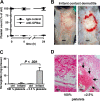
Figure 1
Figure 1
Bleeding in irritant contact dermatitis in thrombocytopenic mice. (A) Thrombocytopenia was achieved by administration of a rat antimouse GPIbα antibody. Platelet counts were determined at indicated time points and displayed in relation to the baseline level (100%). Platelet depletion occurred within minutes (first data point = 0.1 hours) and platelet counts remained < 2.5% for or over 24 hours). Control IgG antibody injection did not alter platelet counts. (B) WT mice were subjected to irritant contact dermatitis (ICD) in the presence and absence of platelets. Erythema and edema were seen in both groups, while massive bleeding into the skin at the inflammatory ring was seen only in thrombocytopenic mice (right). Mice were killed 4 hours after stimulation. Bar = 5 mm. (C) Hemoglobin content in biopsies of the ICD-treated skin. Control skin has barely detectable levels of hemoglobin that were significantly elevated in the inflamed skin of thrombocytopenic mice (P < .001; n = 5). Error bars represent SEM. (D) Skin sections stained with hematoxylin and eosin show massive RBC accumulation (arrows) in the tissue only of ICD-challenged thrombocytopenic animals. Bar = 40 μm.
Figure 2
Figure 2
Hemorrhage at sites of the reverse passive Arthus reaction in platelet-depleted mice. (A) Mice were subjected to rpA (→) in the presence and absence of platelets and killed at 4 hours. Erythema was seen in control mice (left), while massive petechial bleeding was observed only in thrombocytopenic animals (right). Bar = 5 mm. (B) Evans blue leakage into the tissue was measured to assess the size of the inflammatory area. No differences were found between control and platelet-depleted animals. (C) Hemoglobin content in biopsies of the inflamed and control skin. Induction of the rpA, but not the IgG control, led to significantly elevated Hb levels in thrombocytopenic mice (*P < .001; n = 4). Error bars represent SEM. (D) Skin sections stained with H&E revealed accumulation of RBCs (→) in the inflamed skin of thrombocytopenic mice. Bar = 100 μm.
Figure 3
Figure 3
Kinetics of inflammatory bleeding in thrombocytopenic mice during reverse passive Arthus reaction. (A) Photographs of progressing rpA in the dorsal skinfold chamber. In the absence of platelets, petechial bleeding was clearly visible after 2 hours and increased with time. In nondepleted animals there were virtually no petechial spots. Window diameter was 12 mm. (B) Microscopic view of the progressing rpA in a thrombocytopenic mouse. Petechial bleeding was detected at 20 minutes, with further growth of the spot at 40 minutes. Bar = 200 μm. (C) Petechial spots visible to the eye (∼ 100 μm) were counted during rpA in thrombocytopenic and control animals. The difference in incidence of petechiae became statistically significant within 1 hour (P < .01; n = 4). At t = 4 hours, the petechiae became confluent, impairing quantification. Error bars represent SEM.
Figure 4
Figure 4
Rescue of inflammatory hemorrhage by minor platelet transfusion. The rpA model was studied in thrombocytopenic mice receiving transfusion of depletion-resistant platelets from IL4Rα/GPIbα-tg mice. Bars indicate the area of hemorrhage in mice at various platelet counts. Photographs (right) show representative rpA reaction spots. Bar = 5 mm. Transfusion of platelets to 5% to 8% of normal platelet count significantly prevented bleeding (P < .01). At levels of 10% to 15% circulating platelets, the hemorrhagic area was similar to nondepleted control mice (P = .15; n = 5). Error bars represent SEM.
Figure 5
Figure 5
Inflammatory hemorrhage in stroke. Brain sections of mice challenged by the middle cerebral artery occlusion stroke model and analyzed 24 hours after reperfusion. The inflamed hemisphere (right) shows hemorrhagic spots in the thrombocytopenic mice (→).
Figure 6
Figure 6
Lung inflammation induces severe hemorrhage and anemia during thrombocytopenia. (A) Top: Postmortem analysis of lungs after intranasal LPS challenge. Massive pulmonary hemorrhage is observed in platelet-depleted lungs on the right. Bar = 5 mm. Middle: Bronchoalveolar lavage reveals red opaque fluid from depleted (dep) mice and not from control (co) mice with normal platelet count. Bottom: Lung sections stained with hematoxylin and eosin show massive RBC infiltration in airways and alveolar space of thrombocytopenic mice. Bar = 100 μm. (B) Significantly increased amounts of RBCs were found in the lavage of inflamed thrombocytopenic mice (P < .001; n = 5). Platelet depletion per se did not induce RBC loss into the lung. (C) Hemoglobin levels in whole blood of thrombocytopenic and control mice challenged in the lung inflammation model. Pulmonary hemorrhage is accompanied by a significant drop in hemoglobin levels in the thrombocytopenic animals. Error bars represent SEM.

Comment in

References

    1. Massberg S, Schurzinger K, Lorenz M, et al. Platelet adhesion via glycoprotein IIb integrin is critical for atheroprogression and focal cerebral ischemia: an in vivo study in mice lacking glyco-protein IIb. Circulation. 2005;112:1180–1188. - PubMed
    1. Massberg S, Brand K, Gruner S, et al. A critical role of platelet adhesion in the initiation of atherosclerotic lesion formation. J Exp Med. 2002;196:887–896. - PMC - PubMed
    1. Iannacone M, Sitia G, Isogawa M, et al. Platelets mediate cytotoxic T lymphocyte-induced liver damage. Nat Med. 2005;11:1167–1169. - PMC - PubMed
    1. Burger PC, Wagner DD. Platelet P-selectin facilitates atherosclerotic lesion development. Blood. 2003;101:2661–2666. - PubMed
    1. Cambien B, Bergmeier W, Saffaripour S, Mitchell HA, Wagner DD. Antithrombotic activity of TNF-alpha. J Clin Invest. 2003;112:1589–1596. - PMC - PubMed

Publication types