. 2008 May;19(5):923-32.
doi: 10.1681/ASN.2007090982. Epub 2008 Feb 6.

TLR4 signaling mediates inflammation and tissue injury in nephrotoxicity

Affiliations

TLR4 signaling mediates inflammation and tissue injury in nephrotoxicity

Binzhi Zhang et al. J Am Soc Nephrol. 2008 May.

Abstract

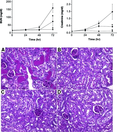
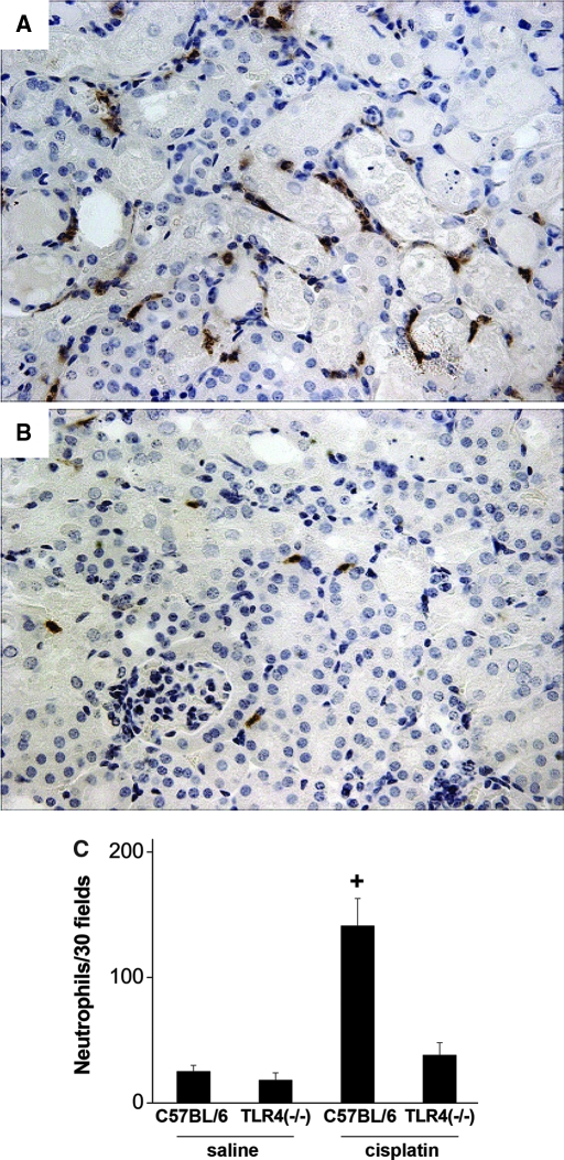
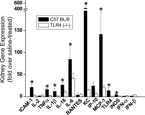
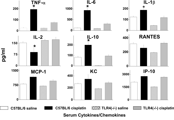
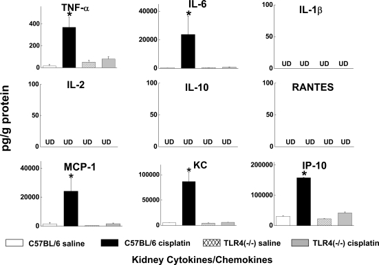
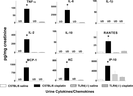
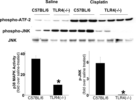
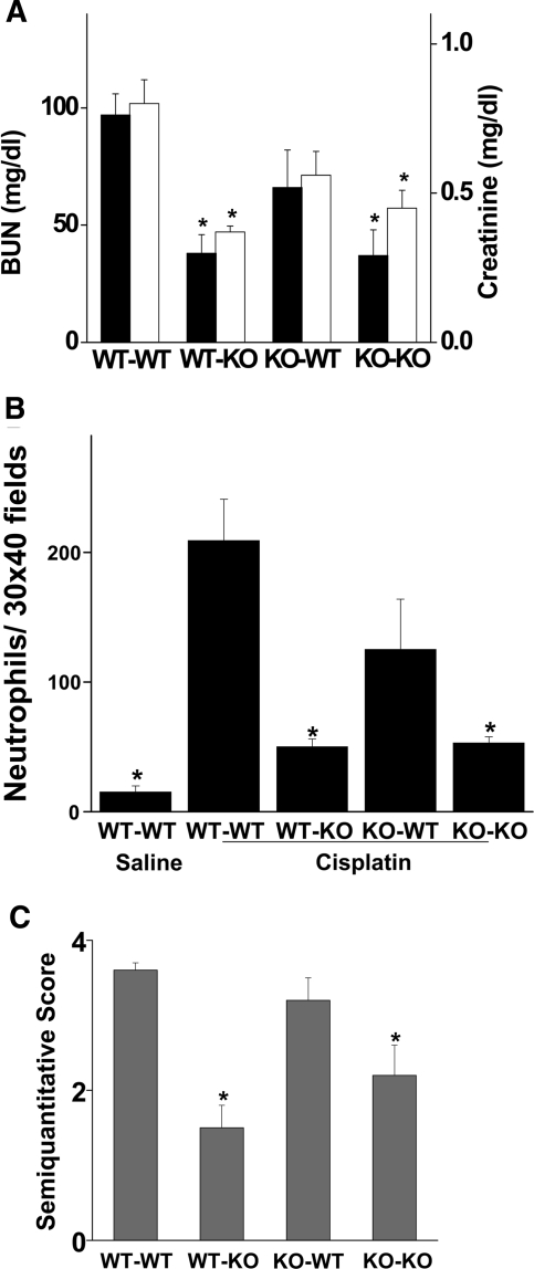
The molecular mechanisms of acute kidney injury (AKI) remain unclear. Toll-like receptors (TLRs), widely expressed on leukocytes and kidney epithelial cells, regulate innate and adaptive immune responses. The present study examined the role of TLR signaling in cisplatin-induced AKI. Cisplatin-treated wild-type mice had significantly more renal dysfunction, histologic damage, and leukocytes infiltrating the kidney than similarly treated mice with a targeted deletion of TLR4 [Tlr4(-/-)]. Levels of cytokines in serum, kidney, and urine were increased significantly in cisplatin-treated wild-type mice compared with saline-treated wild-type mice and cisplatin-treated Tlr4(-/-) mice. Activation of JNK and p38, which was associated with cisplatin-induced renal injury in wild-type mice, was significantly blunted in Tlr4(-/-) mice. Using bone marrow chimeric mice, it was determined that renal parenchymal TLR4, rather than myeloid TLR4, mediated the nephrotoxic effects of cisplatin. Therefore, activation of TLR4 on renal parenchymal cells may activate p38 MAPK pathways, leading to increased production of inflammatory cytokines, such as TNF-alpha and subsequent kidney injury. Targeting the TLR4 signaling pathways may be a feasible therapeutic strategy to prevent cisplatin-induced AKI in humans.


Figures

Figure 1.
Figure 1.
Dependence of cisplatin nephrotoxicity on TLR4. (Top) Effect of cisplatin on renal function in C57BL/6 (▪) or Tlr4(−/−) (•) mice. Male mice were injected with saline (▴) or 20 mg/kg cisplatin (▪, •). Blood urea nitrogen and creatinine were measured at the indicated time points. *P < 0.05 versus C57BL/6 cisplatin-injected mice; n = 4 to 7. (Bottom) Effect of cisplatin on kidney morphology in C57BL/6 (A and C) and Tlr4(−/−) mice (B and D). Kidneys were removed 72 h after injection with cisplatin (A and B) or saline (C and D). Sections were stained with PAS (original magnification ×40). Wild-type mice exhibit severe tubular necrosis, tubular dilation, and cast formation throughout the cortex. Tlr4(−/−) mice have relatively well-preserved renal morphology.
Figure 2.
Figure 2.
Neutrophil infiltration in cisplatin nephrotoxicity requires the presence of TLR4. Sections of kidney harvested from C57BL/6 (A) or Tlr4(−/−) (B) mice 72 h after cisplatin injection were stained for neutrophils as described in “Concise Methods.” (C) Thirty ×40 fields of kidney cortex were examined from each animal. The total number of neutrophils in those 30 fields is presented. +P < 0.01 versus all others; n = 3 to 7.
Figure 3.
Figure 3.
Dependence of renal gene expression on the presence of TLR4. Kidney mRNA abundance of ICAM-1, various cytokines and chemokines, TLR4, and iNOS were determined by real-time RT-PCR. Expression levels are shown as relative fold changes compared with saline-treated mice of the same genotype. *P < 0.05 versus Tlr4(−/−); n = 3.
Figure 4.
Figure 4.
Effects of cisplatin on the serum cytokine expression profile. Serum cytokine levels were measured using a bead-based multiplexed cytokine analysis system. Samples were obtained from C57BL/6 or Tlr4(−/−) mice 72 h after saline or cisplatin injection. *P < 0.05, C57BL/6 versus TLR4(−/−); n = 3 to 5.
Figure 5.
Figure 5.
Effects of cisplatin on kidney cytokine levels. Kidneys were obtained from C57BL/6 or Tlr4(−/−) mice 72 h after saline or cisplatin injection. *P < 0.05 C57BL/6 versus Tlr4(−/−); n = 3 to 5. UD, undetectable.
Figure 6.
Figure 6.
Effects of cisplatin on urine cytokine excretion. Urine samples were obtained from C57BL/6 or Tlr4(−/−) mice 72 h after saline or cisplatin. Cytokine levels were normalized to the creatinine concentration in the urine. *P < 0.05 C57BL/6 versus Tlr4(−/−); n = 3 to 5. UD, undetectable.
Figure 7.
Figure 7.
Effects of cisplatin treatment on p38 MAPK activity and JNK phosphorylation. Kidneys were removed from C57BL/6 or Tlr4(−/−) mice 72 h after injection with either saline or cisplatin and homogenized. p38 MAPK activity was determined by immunoprecipitation and phosphorylation of ATF-2, and JNK phosphorylation was determined by Western blot analysis. Densitometry of results from two independent experiments is shown below the blots. *P < 0.05 versus C57BL/6.
Figure 8.
Figure 8.
Cisplatin induced kidney dysfunction and renal neutrophil infiltration in chimeric mice. (A) Bone marrow chimeric mice were injected with 20 mg/kg cisplatin. Blood urea nitrogen (solid bars) and creatinine (open bars) were measured at 72 h after injection. *P < 0.05 versus WT-WT; n = 3 to 5. (B) Sections of kidney harvested 72 h after injection were stained for neutrophils as described in “Concise Methods.” Thirty ×40 fields of kidney cortex were examined from each animal. The total number of leukocytes in those 30 fields is presented. *P < 0.05 versus WT-WT; n = 3 to 5. (C) Tubular injury was assessed in PAS-stained sections using a semiquantitative scale as described in “Concise Methods.” *P < 0.05 versus WT-WT; n = 3 to 5.
Figure 9.
Figure 9.
Immunofluorescence localization of TLR4 after injection of saline or cisplatin. Representative kidney sections obtained from C57BL/6 (A-D) or Tlr4(−/−) (E and F) mice 48 h after injection. Green fluorescence represents FITC-phalloidin staining of the brush border. Red fluorescence represents TLR4 immunostaining. Blue represents DAPI staining of nuclei. (A) Saline-treated C57BL/6; TLR4 staining is seen in the interstitium and brush borders of proximal tubules (original magnification ×40). (B) A single tubule at higher magnification (original magnification ×100). (C) Cisplatin-treated C57BL/6 (original magnification ×40) shows enhanced brush border staining with clear colocalization of TLR4 and phalloidin. (D) Intracellular TLR4 staining (arrowheads) is also apparent at higher magnification (original magnification ×100). (E-F) Saline-treated Tlr4(−/−) kidneys as a negative control for TLR4 immunostaining.

References

    1. Ries F, Klastersky J: Nephrotoxicity induced by cancer chemotherapy with special emphasis on cisplatin toxicity. Am J Kidney Dis 13: 368–379, 1986 - PubMed
    1. Ramesh G, Reeves WB: TNF-α mediates chemokine and cytokine expression and renal injury in cisplatin nephrotoxicity. J Clin Invest 110: 835–842, 2002 - PMC - PubMed
    1. Ramesh G, Reeves WB: TNFR2-mediated apoptosis and necrosis in cisplatin-induced acute renal failure. Am J Physiol Renal Physiol 54: F610-F618, 2003 - PubMed
    1. Ramesh G, Reeves WB: p38 MAP kinase inhibition ameliorates cisplatin nephrotoxicity in mice. Am J Physiol Renal Physiol 289: F166-F174, 2005 - PubMed
    1. Tsuruya K, Ninomiya T, Tokumoto M, Hirakawa M, Matsutani K, Taniguchi M, Fukuda K, Kanai H, Kishihara K, Hirakata H, Iida M: Direct involvement of the receptor-mediated apoptotic pathways in cisplatin-induced renal tubular cell death. Kidney Int 63: 72–82, 2003 - PubMed

Publication types

MeSH terms