

What makes centromeric cohesion resistant to separase cleavage during meiosis I but not during meiosis II?
- PMID: 18256525
- PMCID: PMC2956405
- DOI: 10.4161/cc.7.2.5325
What makes centromeric cohesion resistant to separase cleavage during meiosis I but not during meiosis II?
Abstract
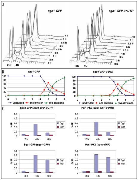
Segregation of chromosomes during meiosis I is triggered by separase cleavage of the cohesin's Rec8 subunit along chromosome arms. Centromeric cohesin is protected from separase cleavage during meiosis I by Sgo1/MEI-S332 proteins in complex with protein phosphatase 2A (PP2A). This retention of centromeric sister chromatid cohesion is essential for faithful segregation of chromatids during the second meiotic division. While Sgo1/PP2A complex is required for protecting centromeric sister chromatid cohesion during meiosis I, it is not known what renders the centromeric cohesion sensitive to separase cleavage during meiosis II. Our data suggest that the absence of Sgo1 and PP2A from meiosis II centromeres is not sufficient to render centromeric cohesion sensitive to cleavage by separase and additional factors are required to ensure the removal of centromeric cohesion during meiosis II.
Figures
References
-
- Page SL, Hawley RS. Chromosome choreography: the meiotic ballet. Science. 2003;301:785–9. - PubMed
-
- Petronczki M, Siomos MF, Nasmyth K. Un menage a quatre: the molecular biology of chromosome segregation in meiosis. Cell. 2003;112:423–40. - PubMed
-
- Ishiguro K, Watanabe Y. Chromosome cohesion in mitosis and meiosis. J Cell Sci. 2007;120:367–9. - PubMed
-
- Lee J, Okada K, Ogushi S, Miyano T, Miyake M, Yamashita M. Loss of Rec8 from chromosome arm and centromere region is required for homologous chromosome separation and sister chromatid separation, respectively, in mammalian meiosis. Cell Cycle. 2006;5:1448–55. - PubMed
-
- Gregan J, Rabitsch PK, Sakem B, Csutak O, Latypov V, Lehmann E, Kohli J, Nasmyth K. Novel genes required for meiotic chromosome segregation are identified by a high-throughput knockout screen in fission yeast. Curr Biol. 2005;15:1663–9. - PubMed
Publication types
MeSH terms
Substances
Grants and funding
LinkOut - more resources
Full Text Sources