. 2008 Feb 20;27(4):606-17.
doi: 10.1038/emboj.2008.9. Epub 2008 Feb 7.

WGEF activates Rho in the Wnt-PCP pathway and controls convergent extension in Xenopus gastrulation

Affiliations

WGEF activates Rho in the Wnt-PCP pathway and controls convergent extension in Xenopus gastrulation

Kosuke Tanegashima et al. EMBO J. .

Abstract

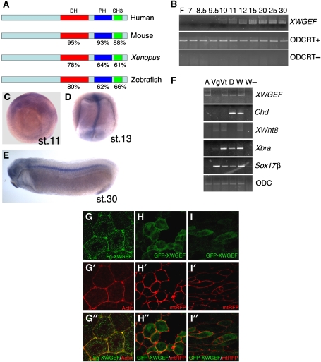
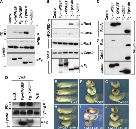
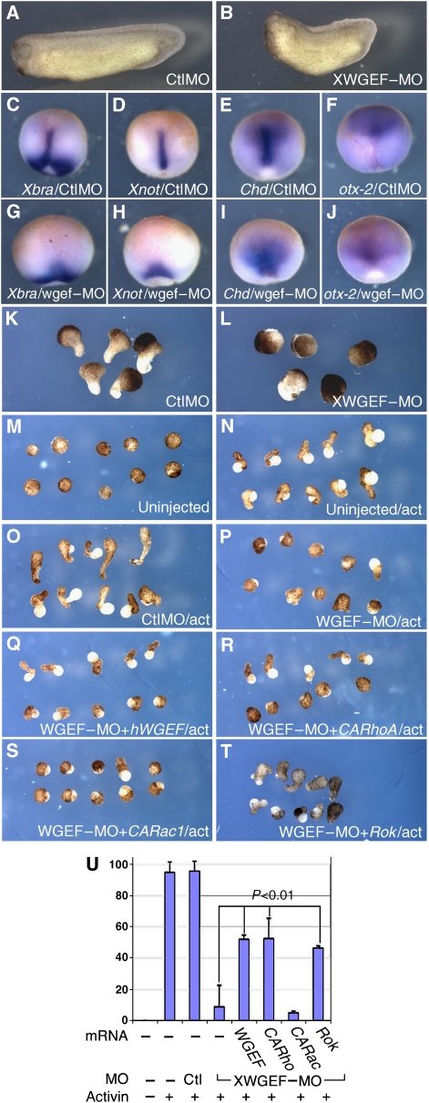
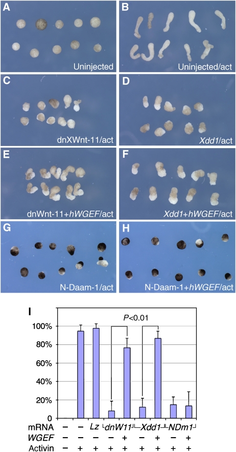
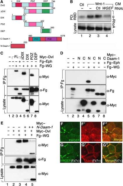
The Wnt-PCP (planar cell polarity, PCP) pathway regulates cell polarity and convergent extension movements during axis formation in vertebrates by activation of Rho and Rac, leading to the re-organization of the actin cytoskeleton. Rho and Rac activation require guanine nucleotide-exchange factors (GEFs), but the identity of the GEF involved in Wnt-PCP-mediated convergent extension is unknown. Here we report the identification of the weak-similarity GEF (WGEF) gene by a microarray-based screen for notochord enriched genes, and show that WGEF is involved in Wnt-regulated convergent extension. Overexpression of WGEF activated RhoA and rescued the suppression of convergent extension by dominant-negative Wnt-11, whereas depletion of WGEF led to suppression of convergent extension that could be rescued by RhoA or Rho-associated kinase activation. WGEF protein preferentially localized at the plasma membrane, and Frizzled-7 induced colocalization of Dishevelled and WGEF. WGEF protein can bind to Dishevelled and Daam-1, and deletion of the Dishevelled-binding domain generates a hyperactive from of WGEF. These results indicate that WGEF is a component of the Wnt-PCP pathway that connects Dishevelled to Rho activation.


Figures

Figure 1
Figure 1
Molecular cloning and expression pattern of XWGEF. (A) Amino-acid sequence comparison of WGEF proteins. The DH, PH and SH3 domains of hWGEF (BC040640) share high sequence identity with mouse (AAH60376, Rho GEF 19), Xenopus (DQ640641, this study) and zebrafish (XP_697662, predicted sequence of Rho GEF 19-related protein) proteins. (B) Developmental expression of XWGEF. RT–PCR analysis was performed at various stages as indicated (Nieuwkoop and Faber, 1956); F, fertilized eggs. (CE) In situ hybridization with XWGEF. (C) Vegetal view of stage-11 embryo showing widespread expression. (D) Dorsal view of stage-13 embryo; preferential expression of XWGEF was detected in the notochord. (E) Lateral view of stage-30 embryo showing XWGEF expression in notochord and head region. (F) RT–PCR with RNA from dissected stage-10 gastrula: animal (A) and vegetal (Vg) regions, and ventral (Vt) and dorsal (D) marginal zone; XWGEF transcripts were present in animal and marginal regions. Chd, Wnt8, Xbra and Sox17β served as markers for dorsal mesoderm, ventral mesendoderm, entire mesoderm and endoderm, respectively. W, whole embryo; W−, whole embryo without reverse transcriptase. (GI) XWGEF is preferentially localized at the plasma membrane. Fg–XWGEF mRNA (50 pg) or GFP–XWGEF (100 pg) and mtRFP (100 pg) mRNA were injected into animal (H) or dorsal blastomeres (G, I) of four-cell-stage embryos. (G–G″) Fg–XWGEF localization. Dorsal (so-called Keller) explants were dissected at stage 10, fixed at mid-gastrula stage and stained with anti-Flag antibody (G) and Texas Red-conjugated phalloidin to visualize F-actin (G′); merged image (G″). Most of XWGEF protein was at the plasma membrane and colocalized with actin. Staining of explants from uninjected embryos with anti-Flag antibody showed no specific staining (data not shown). (H, I) GFP–XWGEF localization. GFP signal was visualized in live explants at mid-gastrula in animal caps (H), or at early neurula in Keller explants (I). mtRFP outlined the cell membranes (H′, I′). The merged images are shown in (H″, I″). GFP–XWGEF showed preferential membrane localization.
Figure 2
Figure 2
WGEF activates RhoA in cultured cells and Xenopus embryos. (A, B) hWGEF and XWGEF induce RhoA but not Rac1 and Cdc42 activation in HEK293T cells. Flag (Fg)-tagged hWGEF, XWGEF, Ephexin (positive control) and an inactive, DH domain-deleted form of hWGEF (ΔGEF; negative control) were transfected into HEK293T cells. Ephexin activates RhoA, Rac1 and Cdc42 (Shamah et al, 2001). (A) GTP–Rho was precipitated using RBD–GST and detected by anti-RhoA antibody. Endogenous RhoA and Flag-tagged GEF proteins in lysates were detected by anti-RhoA and anti-Flag antibody, respectively. (B) GTP-bound Rac1 and Cdc42 were precipitated by GST–PBD and detected by α-Rac1 and α-Cdc42 antibodies. (C) Fg–hWGEF and Fg–XWGEF bind to RhoA. Flag-tagged GEF constructs were transfected into the 293T cells, pulled down with GST–RhoA, Rac1 or Cdc42 and detected with Flag antibody. (D) WGEF activates RhoA in the Xenopus embryo. A 1-ng of Fg–hWGEF or Fg–XWGEF was injected into the ventral region of four-cell-stage embryos and VMZ was dissected at stage 10. (EH) Overexpression of WGEF caused short body axis formation and suppression of head structures. mRNA was injected into the dorsal side of Xenopus embryos at the four-cell stage. (E) Injection of 250 pg of lacZ; (F) 250 pg of Fg–hWGEF; (G) 250 pg of Fg–XWGEF; (H) 250 pg of Fg–XWGEF and 500 pg of dnRhoA (hRhoAN19); (I) 250 pg of Fg–XWGEF and 500 pg of dnRok; (J) 250 pg of Fg–XWGEF and 500 pg of dnRac1 (hRac1N17). Numbers of embryos are given in Table I.
Figure 3
Figure 3
Depletion of XWGEF suppressed CE movements. (A, B) XWGEF–MO suppressed axis elongation. Sixty nanograms of control MO (CtlMO) (A) or XWGEF–MO (B) were injected into dorsal blastomeres at the four-cell stage. XWGEF–MO-injected embryos showed shortened axis (58/62 embryos), whereas CtlMO-injected embryos did not shows shortened axis (0/57). (CJ) XWGEF–MO suppressed elongation of the notochord, but did not inhibit mesodermal marker expression. Sixty nanograms of CtlMO (C–F) or XWGEF–MO (G–J) were injected into dorsal blastomeres at the four-cell stage together with lacZ RNA to mark the site of injection (red). WISH is shown for Xbra (C, G), Xnot (D, H), Chd (E, I), and Otx-2 (F, J) at stage 13. (K, L) Keller sandwich explants from MO-injected embryos. Two dorsal sectors were dissected from stage-10 embryos and combined with each other. Explants injected with 60 ng of CtlMO extended (K, 42/50), whereas explants injected with 60 ng of XWGEF–MO did not extend (L, 4/41). The difference is statistically significant (P=3 × E−13; Supplementary Table S1). (MT) XWGEF–MO inhibited CE in activin-treated animal caps, and this inhibition was rescued by expression of hWGEF, CARhoA or Rok. MOs with or without mRNA were injected into the animal region at the four-cell stage. Animal caps were dissected at stage 9, treated with activin for 3 h and photographed when sibling embryos reached stage 20. No elongation was seen without activin (M, 0/70 explants elongated), but elongation was induced by activin in uninjected (N, 73/77) or CtlMO-injected (60 ng) explants (O, 62/65). Injection of XWGEF–MO (60 ng) inhibited elongation (P, 7/80 elongated), which was rescued by co-injection of 1 or 2 pg of hWGEF mRNA (Q, 43/83), 1 pg of CARhoA mRNA (R, 33/63) or 50 pg of Rok mRNA (T, 19/41), but not CARac1 mRNA (S, 2/61). (U) Bar graph showing the percentage of elongated animal caps in the experiments shown in panels M–T. Standard error bars are shown. Co-injection of WGEF, CARho and Rok mRNA showed statistically significant rescue compared with explants injected with XWGEF–MO alone (P<0.01; Supplementary Table S1).
Figure 4
Figure 4
WGEF acts within the Wnt–PCP pathway. mRNAs were injected into the animal region at the four-cell stage, animal caps were dissected at stage 9, treated with activin and elongation was observed at equivalent stage 20. Animal caps did not elongate without activin (A, 0/54), but did so after activin treatment (B, 102/108). Injection of 1 ng of dnXWnt-11 (C, 4/49) or Xdd1 (D, 5/41) mRNA suppressed elongation, whereas lacZ did not suppress elongation (data not shown; 41/42). (E) Inhibition by dnXWnt-11 was rescued by co-injection of 20 pg of hWGEF mRNA (52/68), but inhibition by Xdd1 was only partially rescued (F, 39/45 explants elongated to a lesser extent). (G) N-Daam-1 (2 ng mRNA) inhibited CE (G; 7/47) and 20 pg of hWGEF mRNA failed to rescue this inhibition (H, 7/52). (I) Bar graph showing the percentage of elongated animal caps in the experiments shown in panels A–H. Standard error bars are shown. P<0.01 for both comparisons (Supplementary Table S1).
Figure 5
Figure 5
WGEF interacts with Wnt–PCP pathway components. (A) Schematic representation of epitope-tagged constructs of Dvl-2 and Daam-1. (B) Depletion of hWGEF blocks Rho activation by Wnt signaling. hWGEF or control (Ctl) RNAi was transfected into MCF-7 cells, and active RhoA was measured. Wnt-1-conditioned media (CM) stimulated the activation of RhoA (lane 2) as compared with Ctl CM (lane 1). Ctl RNAi had no effect (lane 3), but RNAi against hWGEF blocked the activation of Rho above control levels (lane 4). (CE) In co-immunoprecipitation experiments, the antibodies used for precipitation are indicated by IP, and the antibodies used for blotting are shown on the right of each panel. (C) Dvl binds to hWGEF through its PDZ domain. Myc-tagged Dvl-2, Dvl-2ΔDIX and Dvl-2–PDZ co-precipitated with Fg–hWGEF (Fg–WG), but Myc–Dvl-2 did not bind to Fg–Ephexin (Fg–Eph). (D) Myc-tagged N-Daam-1 (N) but not C-Daam-1 (C) co-precipitated with Fg–WG, but neither co-precipitated with Fg–Eph. (E) Myc-tagged Dvl-2 binds to Fg–WG and this binding is abolished in a dose-dependent manner by cotransfection with Myc–N-Daam-1, a dominant-negative form of Daam-1. (F, G) Fz enhances the colocalization of Dsh and WGEF. Fg–XWGEF (50 pg) and Myc–XDsh (250 pg) mRNA was injected into the animal pole with (G) or without (F) 1 ng of XFz-7 mRNA. Animal caps were dissected and stained with Flag (F, G) and Myc (F′, G′) antibody, and photographed using confocal microscopy. Merged images are shown in F″ and G″.
Figure 6
Figure 6
The N-terminal domain of hWGEF binds to Dvl and acts as an autoinhibitory domain. (A) Constructs of hWGEF, all of which were Flag tagged, and summary of Dvl binding. (B) Binding experiments were carried out after transfection into HEK293T cells. Antibodies used for blotting are indicated to the right of each panel. Only constructs that retain the N-terminal domain of hWGEF bind to Dvl-2. (C) Deletion of the Dvl-binding domain generates hyperactive WGEF. Constructs were transfected into HEK293T cells followed by assay for active RhoA. (D) WGEFΔN is more active than wild-type WGEF in Rho activation in the Xenopus embryo. Different constructs (100 pg of RNA) were injected into the VMZ at the four-cell stage, and dissected and assayed at stage 10. At these doses of injected RNA, wild-type hWGEF and XWGEF are not effective in Rho activation, but both N-terminal deletion (ΔN) constructs are strongly active. (E) The N-terminus deleted (ΔN) form of WGEF binds RhoA more effectively than the wild-type protein. In vitro translated FghWGEF, FghWGEFΔN and Fg–hWGEFΔGEF were tested by pull-down assay with RhoA–GST; Fg–hWGEFΔGEF is included as negative control. The ratio of Fg–hWGEFΔN to Fg–hWGEF binding to RhoA–GST was 3.9±0.93; n=4. (FJ) Overexpression of WGEFΔN is more effective than wild-type WGEF in the induction of embryonic defects. RNAs encoding the indicated constructs were injected into the dorsal side of Xenopus embryos at the four-cell stage; the amounts in picograms are indicated. Numbers of embryos are given in Table I. (K, L) A model for the interaction of Wnt–PCP components in regulating CE. (K) In the absence of Wnt signaling, Dvl, Daam-1 and Rho are in the cytosol (Park et al, 2006; Kim and Han, 2007), whereas WGEF is present at the membrane; Rho is not active. (L) Upon Wnt signalling and Fz activation, Dvl, Daam-1 and Rho are recruited to the membrane (Park et al, 2006; Kim and Han, 2007) and come to be colocalized and complexed with WGEF, leading to Rho activation.

Similar articles

Cited by

References

    1. Asashima M, Nakano H, Simada K, Ishii K, Shibai H, Ueno N (1990) Mesodermal induction in early amphibian embryos by activin A (erythroid differentiation factor). Rouxs Arch Dev Biol 198: 330–335 - PubMed
    1. Benard V, Bohl BP, Bokoch GM (1999) Characterization of rac and cdc42 activation in chemoattractant-stimulated human neutrophils using a novel assay for active GTPases. J Biol Chem 274: 13198–13204 - PubMed
    1. Cheyette BN, Waxman JS, Miller JR, Takemaru K, Sheldahl LC, Khlebtsova N, Fox EP, Earnest T, Moon RT (2002) Dapper, a dishevelled-associated antagonist of beta-catenin and JNK signaling, is required for notochord formation. Dev Cell 4: 449–461 - PubMed
    1. Copp AJ, Greene ND, Murdoch JN (2003) The genetic basis of mammalian neurulation. Nat Rev Genet 4: 784–793 - PubMed
    1. Crespo P, Schuebel KE, Ostrom AA, Gutkind JS, Bustelo XR (1997) Phosphotyrosine-dependent activation of Rac-1 GDP/GTP exchange by the vav proto-oncogene product. Nature 385: 169–172 - PubMed

Publication types