. 2008 Mar 5;27(5):736-47.
doi: 10.1038/emboj.2008.17. Epub 2008 Feb 7.

Interactions between UPF1, eRFs, PABP and the exon junction complex suggest an integrated model for mammalian NMD pathways

Affiliations

Interactions between UPF1, eRFs, PABP and the exon junction complex suggest an integrated model for mammalian NMD pathways

Pavel V Ivanov et al. EMBO J. .

Abstract

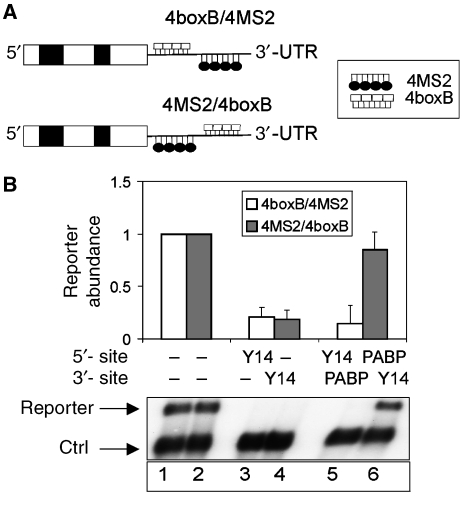
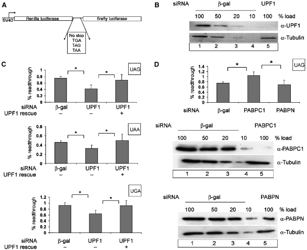
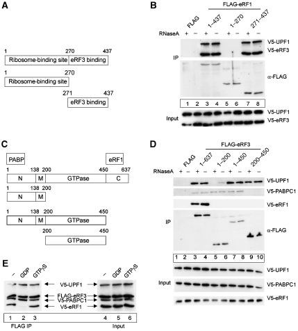
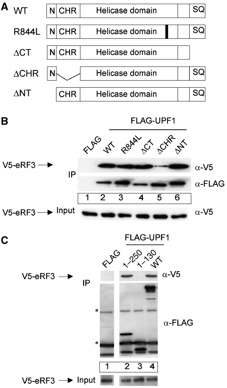
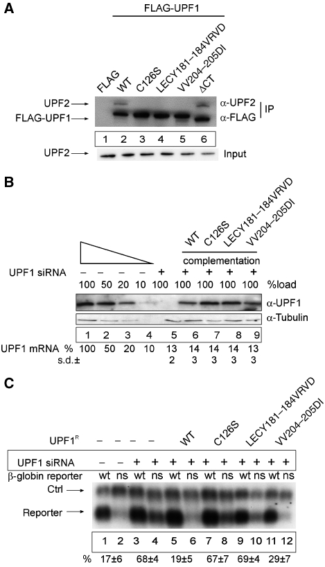
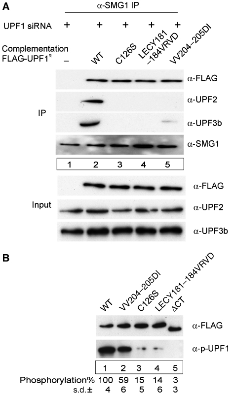
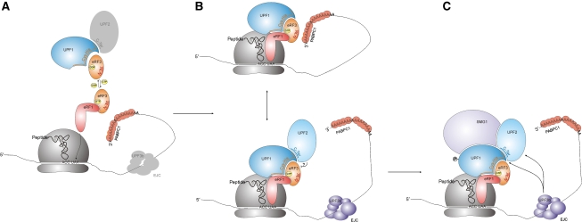
Nonsense-mediated mRNA decay (NMD) represents a key mechanism to control the expression of wild-type and aberrant mRNAs. Phosphorylation of the protein UPF1 in the context of translation termination contributes to committing mRNAs to NMD. We report that translation termination is inhibited by UPF1 and stimulated by cytoplasmic poly(A)-binding protein (PABPC1). UPF1 binds to eRF1 and to the GTPase domain of eRF3 both in its GTP- and GDP-bound states. Importantly, mutation studies show that UPF1 can interact with the exon junction complex (EJC) alternatively through either UPF2 or UPF3b to become phosphorylated and to activate NMD. On this basis, we discuss an integrated model where UPF1 halts translation termination and is phosphorylated by SMG1 if the termination-promoting interaction of PABPC1 with eRF3 cannot readily occur. The EJC, with UPF2 or UPF3b as a cofactor, interferes with physiological termination through UPF1. This model integrates previously competing models of NMD and suggests a mechanistic basis for alternative NMD pathways.


Figures

Figure 1
Figure 1
Antagonistic relationship of PABPC1 and the EJC in stop codon definition. (A) Schematic representation of the 4boxB/4MS2 and 4MS2/4boxB reporters. White and black boxes represent exons and introns of the β-globin gene, respectively. BoxB and MS2 sites are shown as white boxes or black loops, respectively. (B) Tethering of PABPC1 and the EJC protein Y14. HeLa cells were transfected with the indicated 4boxB/4MS2 or 4MS2/4boxB constructs and the plasmids expressing the MS2–PABPC1 and λNV5–Y14 fusion proteins as indicated. Total RNA samples were analysed by northern blotting. The levels of the 4boxB/4MS2 and the 4MS2/4boxB reporter mRNAs were normalized to the control β-globin reporter (ctrl). For each reporter, mRNA levels of the reporter without any tethered factor (MS2 protein and λN peptide alone) were set as 1.0 (lanes 1 and 2). Values were calculated from three independent experiments, error bars represent standard deviations.
Figure 2
Figure 2
Effect of UPF1 and PABPC1 on translational readthrough and termination. (A) Schematic representation of the dual luciferase reporters used for measuring translation termination readthrough. (B, C) UPF1 knockdown decreases the level of readthrough at all three stop codons. Protein lysates from HeLa cells that were transfected with siRNAs against β-galactosidase (β-gal) (B, lanes 1–4) or UPF1 (B, lane 5) were immunoblotted with an anti-UPF1 antibody. A dilution series corresponding to 50, 20 and 10% of the protein amount that was used in lane 1 (20 μg) was loaded to assess the efficiency of UPF1 depletion. Reprobing with a tubulin-specific antibody was performed as a loading control. At 48 h after siRNA depletion, HeLa cells were transfected with the dual luciferase construct (p2luc-stop). The percentage of readthrough at the three different stop codons is shown (C). The results obtained with the construct without a stop codon were set as 100% readthrough. To complement the UPF1 depletion, cells were transfected with 0.2 μg of siRNA-resistant FLAG-UPF1R. The values were calculated from six independent depletion experiments, error bars represent standard deviations. *Statistical significance at P<0.05. (D) PABPC1 knockdown increases the level of readthrough at a UAG stop codon. Depletions of PABPC1 and nuclear PABP were achieved by siRNA treatment as described in (B). HeLa cells were transfected with the dual luciferase construct p2luc-TAG or with the construct without an interposed stop codon p2luc-if 48 hours after siRNA treatment. The percentage of readthrough at the UAG stop codon was calculated as described in (C). *Statistical significance at P<0.05.
Figure 3
Figure 3
Definition of biochemical interactions between UPF1, release factors and PABPC1. (A, B) UPF1 and eRF3 interact with the C-terminal part of eRF1. (A) Schematic representation of the domain structure of human release factor eRF1. The ribosome- and the eRF3-binding domain are depicted. (B) Co-immunoprecipitation experiments with eRF1 fragments. HeLa cells were co-transfected with an empty FLAG plasmid (lanes 1 and 2), with FLAG-eRF1 (lanes 3 and 4) or the indicated FLAG-tagged fragments of eRF1 (lanes 5–8) together with a plasmid for V5-UPF1 and V5-eRF3. Immunoprecipitations were carried out in the presence or absence of RNaseA. V5-tagged proteins were detected by immunoblotting with an anti-V5 antibody. Lysate (5%) used for the immunoprecipitations was loaded in the input lanes. (C, D) Interaction of eRF3 with eRF1, UPF1 and PABPC1. (C) Schematic representation of the domain structure of the human release factor eRF3. N-terminal (N; PABPC1 binding), middle (M), GTPase- and C-terminal (C; eRF1 binding) parts of eRF3 are indicated. (D) Co-immunoprecipitation experiments with eRF3 fragments. HeLa cells were transfected with an empty FLAG plasmid (lanes 1 and 2), with FLAG-eRF3 (lanes 3 and 4) or the indicated FLAG-tagged fragments of eRF3 (lanes 5–10) together with a plasmid for V5-UPF1, V5-PABPC1 and V5-eRF1. Immunoprecipitations were carried out as described in (B). V5-tagged proteins were detected by immunoblotting with an anti-V5 antibody. Lysate (5%) used for the immunoprecipitations was loaded in the input lanes. (E) UPF1 interacts with eRF3 in the presence of GDP or GTP. Co-immunoprecipitation experiments with eRF3 in the presence of GTP or GDP. HeLa cells were transfected with the plasmid FLAG-eRF3 (lanes 1–6) and with plasmids V5-UPF1, V5-PABPC1 and V5-eRF1, respectively. Immunoprecipitation was carried out in the presence of RNaseA and the guanine nucleotides (1 μM) as indicated (lanes 2 and 3). V5-tagged proteins were detected by immunoblotting with an anti-V5 antibody as described above. Lysate (2.5%) used for the immunoprecipitations was loaded in the input lanes (lanes 4–6).
Figure 4
Figure 4
eRF3 binds to the cysteine–histidine-rich region (CHR) of UPF1. (A) Schematic representation of the domain structure of human UPF1. Abbreviations used: WT—wild-type UPF1; R844L—dominant-negative point mutant of the helicase domain; ΔCT—mutant lacking the C-terminal amino acids 1074–1118; ΔCHR—mutant lacking amino acids 130–250 including the CHR; ΔNT—mutant lacking the N-terminal amino acids 1–40 (N); SQ—serine/threonine-glutamine-rich ((S/T)-Q-rich) motifs. (B) Co-immunoprecipitation experiments with UPF1 mutants. HeLa cells were transfected with an empty FLAG plasmid (lane 1), with wild-type FLAG-UPF1 (lane 2), the point mutant FLAG-R844L (lane 3) or the indicated FLAG-tagged fragments of UPF1 (lanes 4–6) together with a V5-eRF3 plasmid. Immunoprecipitations were carried out as described in Figure 3B in the presence of RNaseA. V5-eRF3 was detected by immunoblotting with an anti-V5 antibody. The input lane shows 4% of the lysate used per assay. (C) Co-immunoprecipitation experiments with the N-terminal fragment of UPF1. HeLa cells were transfected with an empty FLAG plasmid (lane 1), with FLAG-UPF1 (lane 4) or the indicated FLAG-tagged fragments of UPF1 (lanes 2 and 3) together with a V5-eRF3 plasmid. Immunoprecipitation and the detection of V5-eRF3 were carried out as described in (B). *Immunoglobulin heavy and light chains. Lysate (5%) used for the immunoprecipitations was loaded in the input lane.
Figure 5
Figure 5
An UPF1 mutant that triggers NMD but fails to interact with UPF2. (A) Mutational analysis of the UPF2-binding site within the UPF1 CHR region. HeLa cells were transfected with empty FLAG plasmid (lane 1), wild-type FLAG-UPF1 (lane 2) or with the indicated FLAG-tagged mutants of UPF1 (lanes 3–6). Cell extract preparation, immunoprecipitation procedure and immunoblotting were carried out as described in Figure 3B in the presence of RNaseA. The UPF2 protein was detected by immunoblotting with an anti-UPF2 antibody. Lysate (5%) used for the immunoprecipitations was loaded in the input lane. (B, C) Functional analysis of UPF1 CHR mutants. (B) Functional complementation of UPF1 depletion. Immunoblotting analysis of lysates obtained from UPF1-depleted and UPF1-complemented cells was performed as described in Figure 2B and as described previously (Gehring et al, 2005). For the complementation of UPF1-depleted cells, cells were transfected with 0.2 μg FLAG-UPF1R. UPF1 mRNA levels were determined by qRT–PCR using primers that are specific for the endogenous mRNA and do not amplify the plasmid-derived UPF1 mRNA. The values were calculated from four independent experiments with standard deviations (±s.d.). (C) Northern blot analysis of RNA isolated from HeLa cells transfected with siRNA against β-galactosidase (lanes 1–2) or UPF1 (lanes 3–12). At 48 h after siRNA transfections, the cells were co-transfected with plasmids for the transfection efficiency control (ctrl), the NMD reporters (reporter; wt—wild-type β-globin, ns—NS39 β-globin mutant) and plasmids expressing the depicted FLAG-UPF1R variants. The values were calculated from three independent experiments. Error bars represent s.d.
Figure 6
Figure 6
SMG1-dependent phosphorylation of UPF1 correlates with binding of UPF2 or UPF3b. (A) SMG1 precipitates UPF1 mutants. Complementation of the UPF1 depletion with the indicated Flag-UPF1R variants was carried out as described in Figure 5B. An anti-SMG1 antibody was used to immunoprecipitate SMG1-complexes. Anti-FLAG, anti-UPF2, anti-UPF3b and anti-SMG1 antibodies were used to visualize the respective proteins in the immunoprecipitates. Lysate (10%) used for the immunoprecipitations was loaded in the input lanes. (B) Mutations in the Cys–His-rich region of UPF1 affect its phosphorylation. HeLa cells were transfected with plasmids expressing the indicated FLAG-UPF1 mutants. After 48 h, cells were lysed and UPF1 mutants were immunoprecipitated with anti-FLAG agarose beads in the presence of phosphatase inhibitors. Immunoprecipitates were extensively washed, bound proteins were eluted and equal amounts of the eluted proteins were separated by SDS–PAGE. Immunoblotting was performed with an anti-phospho-UPF1 antibody (α-p-UPF1) or a FLAG antibody. The bands were quantified using an Odyssey Infrared Imaging System (LI-COR Biosciences).
Figure 7
Figure 7
An integrated model for mammalian NMD pathways. Schematic diagram of the structural and the functional interaction networks between release factors, UPF proteins, PABPC1, SMG1 and the EJC. Note that the diagram is not time-resolved, and hence some depicted interactions may not occur simultaneously and/or be competitive. (A) The Cys–His-rich domain of UPF1 (Cys–His) binds to the C terminus of UPF2 (see also Kadlec et al, 2006) and to the GTPase domain of eRF3. In the presence of GTP (reflecting the pre-termination state), the C-terminal end of eRF1 binds to the C-terminal end of eRF3, whereas this interaction does not occur in the presence of GDP (reflecting the post-termination state). The C terminus of eRF1 may directly bind to UPF1 or this interaction may be bridged by eRF3. (B) The C terminus of PABPC1 binds to the N terminus of eRF3 (N-Ter). During translation termination, UPF1 monitors the correct position of the termination codon. An interaction between PABPC1 and eRF3 ‘certifies' a normal termination event (top panel), whereas a downstream EJC is indicating premature termination (bottom panel). (?)—a possible competition between UPF2 and eRF3 for UPF1 binding. (C) In case of premature termination, the EJC triggers the SMG1-dependent UPF1 phosphorylation through UPF2 or UPF3b, which leads to NMD.

Similar articles

Cited by

References

    1. Alkalaeva EZ, Pisarev AV, Frolova LY, Kisselev LL, Pestova TV (2006) In vitro reconstitution of eukaryotic translation reveals cooperativity between release factors eRF1 and eRF3. Cell 125: 1125–1136 - PubMed
    1. Altamura N, Groudinsky O, Dujardin G, Slonimski PP (1992) NAM7 nuclear gene encodes a novel member of a family of helicases with a Zn-ligand motif and is involved in mitochondrial functions in Saccharomyces cerevisiae. J Mol Biol 224: 575–587 - PubMed
    1. Amrani N, Ganesan R, Kervestin S, Mangus DA, Ghosh S, Jacobson A (2004) A faux 3′-UTR promotes aberrant termination and triggers nonsense-mediated mRNA decay. Nature 432: 112–118 - PubMed
    1. Amrani N, Dong S, He F, Ganesan R, Ghosh S, Kervestin S, Li C, Mangus DA, Spatrick P, Jacobson A (2006) Aberrant termination triggers nonsense-mediated mRNA decay. Biochem Soc Trans 34: 39–42 - PubMed
    1. Andjelkovic N, Zolnierowicz S, Van Hoof C, Goris J, Hemmings BA (1996) The catalytic subunit of protein phosphatase 2A associates with the translation termination factor eRF1. EMBO J 15: 7156–7167 - PMC - PubMed

Publication types