

CT scanning for diagnosing blunt ureteral and ureteropelvic junction injuries
- PMID: 18257927
- PMCID: PMC2258295
- DOI: 10.1186/1471-2490-8-3
CT scanning for diagnosing blunt ureteral and ureteropelvic junction injuries
Abstract
Background: Blunt ureteral and ureteropelvic (UPJ) injuries are extremely rare and very difficult to diagnose. Many of these injuries are missed by the initial trauma evaluation.
Methods: Trauma registry data was used to identify all blunt trauma patients with ureteral or UPJ injuries, from 1 April 2001 to 30 November 2006. Demographics, injury information and outcomes were determined. Chart review was then performed to record initial clinical and all CT findings.
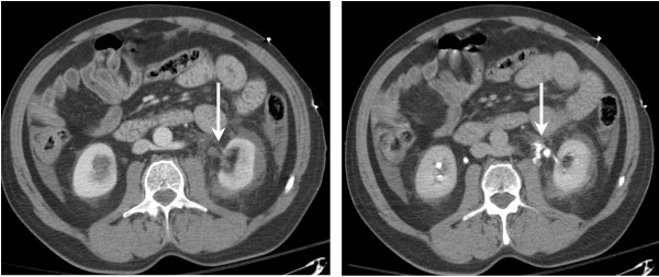
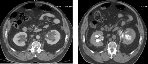
Results: Eight patients had ureteral or UPJ injuries. Subtle findings such as perinephric stranding and hematomas, and low density retroperitoneal fluid were evident on all initial scans, and prompted delayed excretory scans in 7/8 cases. As a result, ureteral and UPJ injuries were diagnosed immediately for these seven patients. These findings were initially missed in the eighth patient because significant associated visceral findings mandated emergency laparotomy. All ureteral and UPJ injuries have completely healed except for the case with the delay in diagnosis.
Conclusion: Most blunt ureteral and UPJ injuries can be identified if delayed excretory CT scans are performed based on initial CT findings of perinephric stranding and hematomas, or the finding of low density retroperitoneal fluid.
Figures
References
-
- Kawashima A, Sandler CM, Corriere JN, Rodgers BM, Goldman SM. Ureteropelvic Junction Injuries Secondary to Blunt Abdominal Trauma. Radiology. 1997;205:487–492. - PubMed
MeSH terms
LinkOut - more resources
Full Text Sources
Medical
Research Materials
Miscellaneous
