

Review
doi: 10.3201/eid1401.070163.
Human ophthalmomyiasis interna caused by Hypoderma tarandi, Northern Canada
Affiliations
- PMID: 18258079
- PMCID: PMC2600172
- DOI: 10.3201/eid1401.070163
Item in Clipboard
Review
Human ophthalmomyiasis interna caused by Hypoderma tarandi, Northern Canada
Emerg Infect Dis.
2008 Jan.
Abstract
Human myiasis caused by bot flies of nonhuman animals is rare but may be increasing. The treatment of choice is laser photocoagulation or vitrectomy with larva removal and intraocular steroids. Ophthalmomyiasis caused by Hypoderma spp. should be recognized as a potentially reversible cause of vision loss.
Figures
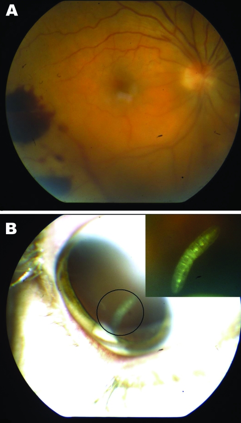
A) Retinal hemorrhages visible on funduscopic examination of right eye of a 41-year-old woman, Nunavut, Canada, with ophthalmomyiasis intern). B) Segmented 3-mm larva with a cylindrical body, no visible spines, and indistinguishable anterior and posterior ends in the vitreous cavity, corresponding to the first instar of Hypoderma tarandi.
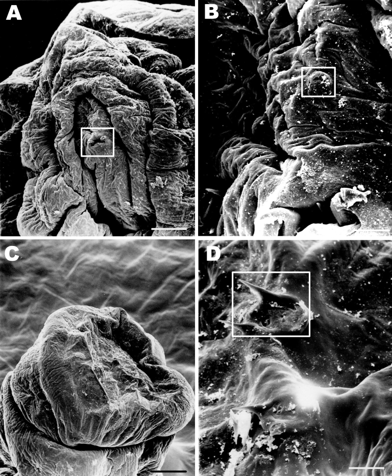
Scanning electron microscope images of the parasite from an 11-year-old Inuit boy, Nunavut, Canada. A) Anterior end of the maggot. The cephalic segment is evident; mouth and mouth hooks are present (boxed). Scale bar = 50 μm. B) The characteristic cephalic sensory array (boxed). Scale bar = 10 μm. C) Posterior segments of the maggot. Scale bar = 100 μm. D) Spiracular openings on the posterior segments of the maggot characteristic of first instar of Hypoderma. Scale bar = 10 μm.
References
-
- Anderson JR. Oestrid myiasis of humans. In: Colwell DD, Hall MJ, Scholl PJ, editors. The oestrid flies—biology, host-parasite relationships, impact and management. Oxford (UK): CABI Publishing; 2006. p. 359.
-
- Custis PH, Pakalnis VA, Klintworth GK, Anderson WB Jr, Machemer R. Posterior internal ophthalmomyiasis. Identification of a surgically removed Cuterebra larva by scanning electron microscopy. Ophthalmology. 1983;90:1583–90. - PubMed
Publication types
MeSH terms
LinkOut - more resources
Full Text Sources
Medical
Miscellaneous