. 2008 Apr 4;283(14):9177-86.
doi: 10.1074/jbc.M707247200. Epub 2008 Feb 7.

Neph1, a component of the kidney slit diaphragm, is tyrosine-phosphorylated by the Src family tyrosine kinase and modulates intracellular signaling by binding to Grb2

Affiliations

Neph1, a component of the kidney slit diaphragm, is tyrosine-phosphorylated by the Src family tyrosine kinase and modulates intracellular signaling by binding to Grb2

Yutaka Harita et al. J Biol Chem. .

Abstract

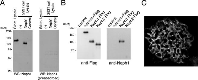
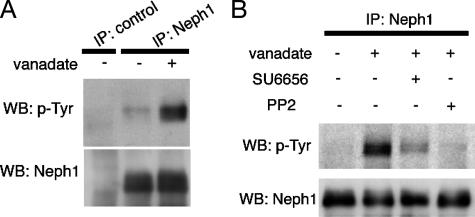
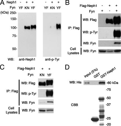
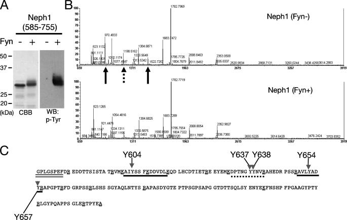
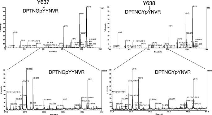
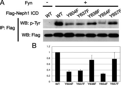
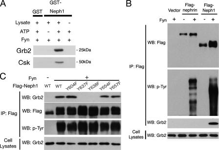
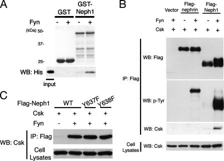
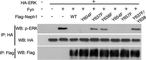
There are several lines of evidence that the podocyte slit diaphragm (SD), which serves as a structural framework for the filtration barrier in kidney glomerulus, also plays an essential role as a signaling platform. Several SD components including nephrin and TRPC6 are known to be phosphorylated by a Src family tyrosine kinase (SFK), Fyn. Here we have characterized Neph1, another SD component, as a novel substrate of SFK. Fyn interacts with and phosphorylates the cytoplasmic domain of Neph1 in vitro and in intact cells. Peptide mass fingerprinting and site-directed mutagenesis identified several tyrosine phosphorylation sites. In pull-down assays using rat glomerular lysates, Neph1 but not nephrin specifically binds to adaptor protein Grb2 and tyrosine kinase Csk in a phosphorylation-dependent manner. Both tyrosine 637 and 638 of Neph1 are crucial for Neph1-Grb2 binding. Phosphorylation of tyrosine 637 is significantly up-regulated in in vivo models of podocyte injury. Furthermore, Neph1 attenuates ERK activation elicited by Fyn, and this inhibitory effect requires the intact binding motif for the Grb2 SH2 domain. Our results shown here demonstrate that Neph1 is a novel in vivo substrate of SFK and suggest that Neph1 modulates ERK signaling through phosphorylation-dependent interaction with Grb2. Thus, SFK orchestrates a wide spectrum of protein-protein interactions and intracellular signaling networks at SD through tyrosine phosphorylation.


Figures

FIGURE 1.
FIGURE 1.
Detection of Neph1 with the anti-Neph1C antibody. A, lysates from isolated rat glomeruli, untransfected 293T cells, 293T cells transiently transfected with plasmid encoding Neph1, or a control vector were separated by SDS-PAGE (10%), transferred to nitrocellulose, and immunoblotted with anti-Neph1C (left panel) or anti-Neph1C preabsorbed with the peptide used for immunization (right panel). B, lysates from 293T cells transiently transfected with plasmid encoding FLAG-tagged Neph1, Neph2, or nephrin were immunoprecipitated by anti-FLAG antibody, and immunoprecipitates were immunoblotted with anti-FLAG or anti-Neph1C antibody. C, indirect immunofluorescence microscopy was performed to detect Neph1 in adult rat kidney cryosections with anti-Neph1C antibody. This antibody specifically labels glomerular podocytes.
FIGURE 2.
FIGURE 2.
Tyrosine phosphorylation of endogenous Neph1 in pervanadate-treated cultured podocytes. A, lysates from cultured podocytes treated with or without 1 mm pervanadate for 15 min were immunoprecipitated with anti-Neph1C antibody, and the immunoprecipitates were immunoblotted with anti-Neph1C or anti-phosphotyrosine (p-Tyr) antibody. B, where indicated, cells were pretreated with 5 μm SU6656 for 60 min or 10 μm PP2 for 15 min prior to treatment with pervanadate. Anti-Neph1C immunoprecipitates (IP) were immunoblotted with anti-phosphotyrosine or anti-Neph1C antibody. WB, Western blot.
FIGURE 3.
FIGURE 3.
Neph1 is bound to and phosphorylated by the Src family tyrosine kinase Fyn. A, 293T cells were transfected with the indicated plasmids encoding an active (YF) or inactive (KN) form of Fyn together with wild-type Neph1. Cell lysates were analyzed by immunoblotting with anti-Neph1C antibody or anti-phosphotyrosine (p-Tyr) antibody. B, 293T cells were transfected with the indicated plasmids, and anti-FLAG immunoprecipitates (IP) or cell lysates were analyzed by immunoblotting with the indicated antibodies. C, 293T cells were transfected with Neph1-Flag together with either the inactive or active form of Fyn. Anti-FLAG immunoprecipitates and cell lysates were analyzed by immunoblotting with antibodies as indicated. D, Fyn binds to Neph1 in vitro. GST or GST-tagged Neph1 cytoplasmic domain (GST-Neph1CD; amino acids 585–755) were immobilized on glutathione beads, and phosphorylated by His-tagged Fyn (active form) in vitro. GST pull-downs were immunoblotted with anti-His antibody. WB, Western blot.
FIGURE 4.
FIGURE 4.
Tyrosine phosphorylation of Neph1 by Fyn in vitro. A, the Neph1 cytoplasmic domain was incubated with or without Fyn in vitro, and the samples were immunoblotted with anti-phosphotyrosine antibody. B, identification of tyrosine residues of Neph1 phosphorylated by Fyn. The same samples as in A were digested with trypsin, and the peptides were analyzed by a MALDI-TOF mass spectrometer. A marked decrease of peak intensity was observed for two peptides (indicated by arrows; 970.5 and 1500.7 Da), and a moderate decrease for another peptide (1198.5 Da, dashed arrow). C, tyrosine phosphorylation sites in the Neph1 cytoplasmic domain. The amino acid sequence of the Neph1-cytoplasmic domain (585–755) with the amino-terminal linker sequence (indicated with a double underline). Peptides corresponding to 970.5 and 1500.7 Da are underlined. A peptide corresponding to 1198.5 Da is indicated by a dashed underline. Candidate phosphorylation sites are indicated by arrows with amino acid numbers. Lysine and arginine residues are marked with single underlines. CBB, Coomassie Brilliant Blue; WB, Western blot.
FIGURE 5.
FIGURE 5.
Identification of tyrosine residues of Neph1 phosphorylated by Fyn by LC-MS/MS analysis. The Neph1 cytoplasmic domain phosphorylated by Fyn was digested with trypsin, and then the peptides were analyzed by offline nanoLC-MALDI-TOF/TOF analysis. The results shown in this figure reveals tyrosine phosphorylation of Neph1 at Tyr637 and Tyr638.
FIGURE 6.
FIGURE 6.
Several tyrosine residues of Neph1 are phosphorylated in intact cells. A, the FLAG-tagged wild-type (WT) or mutant Neph1 cytoplasmic region were transfected into 293T cells together with active Fyn. Cell lysates were immunoprecipitated (IP) with anti-FLAG antibody and immunoprecipitates were immunoblotted for phosphotyrosine. As a control, wild-type Neph1 was transfected without Fyn. B, the densitometry of A is shown. Values are normalized to wild-type Neph1.
FIGURE 7.
FIGURE 7.
Grb2 specifically binds to phosphorylated Neph1. A, recombinant GST-Neph1CD was bound to glutathione-Sepharose beads, and incubated with recombinant Fyn with or without ATP. After washing, the beads were incubated with glomerular lysates from normal rats, and bound proteins were analyzed by immunoblotting for Grb2 and Csk. B, Grb2 binds to phosphorylated Neph1 in 293T cells. 293T cells were transfected with the indicated vectors, and anti-FLAG immunoprecipitates (IP) and cell lysates were analyzed by Western blotting (WB) for FLAG tag, phosphotyrosine, and Grb2. C, both Tyr637 and Tyr638 are required for binding to Grb2. FLAG-tagged wild-type or the indicated mutant Neph1 were transfected into 293T cells together with Fyn, and anti-FLAG immunoprecipitates and cell lysates were analyzed by Western blotting for FLAG tag, phosphotyrosine, and Grb2.
FIGURE 8.
FIGURE 8.
The Csk SH2 domain directly binds to phosphorylated Neph1. A, recombinant GST or GST-Neph1CD were bound to glutathione-Sepharose beads, and incubated with or without Fyn. After washing, the beads were incubated with recombinant His-tagged SH2 domain of Csk, and bound proteins were immunoblotted with anti-His antibody. B, 293T cells were transfected with the indicated vectors, and anti-FLAG immunoprecipitates and cell lysates were analyzed by Western blotting (WB) for FLAG tag, phosphotyrosine (p-Tyr), and Csk. C, 293T cells were transfected with the indicated vectors, and anti-FLAG immunoprecipitates (IP) and cell lysates were analyzed by Western blotting for Csk.
FIGURE 9.
FIGURE 9.
Suppression of Fyn-induced activation of ERK by Neph1. 293T cells were co-transfected with hemagglutinin (HA)-tagged ERK expression vector and a Fyn expression vector together with an empty expression vector, a wild-type Neph1, or each of mutant Neph1 vectors. Anti-hemagglutinin or anti-FLAG immunoprecipitates (IP) were analyzed by Western blotting (WB) with the indicated antibodies.
FIGURE 10.
FIGURE 10.
Tyr637 is phosphorylated in injured podocytes in vivo. A, Neph1 is transiently tyrosine-phosphorylated during foot process effacement in the protamine sulfate-induced podocyte injury model. Rat kidneys were perfused with protamine sulfate for 20 min as described under “Experimental Procedures.” Anti-Neph1C immunoprecipitates (IP) from control or protamine sulfate-treated glomeruli were subjected to immunoblotting with anti-Neph1C or anti-phosphotyrosine antibody. B, recombinant wild-type, Y637F, or Y657F GST-Neph1CD was incubated in vitro with recombinant Fyn, followed by immunoblotting with affinity purified rabbit polyclonal anti-phospho-Neph1 antibody (anti-pY637). C, rat kidneys were perfused with protamine sulfate as in A. Glomerular lysate was immunoprecipitated by anti-Neph1C antibody and the immunoprecipitates were resolved by SDS-PAGE and immunoblotted with anti-Neph1C or anti-phospho-Neph1. D, immunoblot analysis of glomerular lysates from normal and PAN-treated rat kidneys probed with anti-phospho-Neph1. WB, Western blot.

Similar articles

Cited by

References

    1. Pavenstadt, H., Kriz, W., and Kretzler, M. (2003) Physiol. Rev. 83 253-307 - PubMed
    1. Kestila, M., Lenkkeri, U., Mannikko, M., Lamerdin, J., McCready, P., Putaala, H., Ruotsalainen, V., Morita, T., Nissinen, M., Herva, R., Kashtan, C. E., Peltonen, L., Holmberg, C., Olsen, A., and Tryggvason, K. (1998) Mol. Cell. 1 575-582 - PubMed
    1. Putaala, H., Soininen, R., Kilpelainen, P., Wartiovaara, J., and Tryggvason, K. (2001) Hum. Mol. Genet. 10 1-8 - PubMed
    1. Donoviel, D. B., Freed, D. D., Vogel, H., Potter, D. G., Hawkins, E., Barrish, J. P., Mathur, B. N., Turner, C. A., Geske, R., Montgomery, C. A., Starbuck, M., Brandt, M., Gupta, A., Ramirez-Solis, R., Zambrowicz, B. P., and Powell, D. R. (2001) Mol. Cell. Biol. 21 4829-4836 - PMC - PubMed
    1. Roselli, S., Heidet, L., Sich, M., Henger, A., Kretzler, M., Gubler, M. C., and Antignac, C. (2004) Mol. Cell. Biol. 24 550-560 - PMC - PubMed

Publication types

MeSH terms

Substances