

Increased maternofetal calcium flux in parathyroid hormone-related protein-null mice
- PMID: 18258656
- PMCID: PMC2375733
- DOI: 10.1113/jphysiol.2007.149104
Increased maternofetal calcium flux in parathyroid hormone-related protein-null mice
Abstract
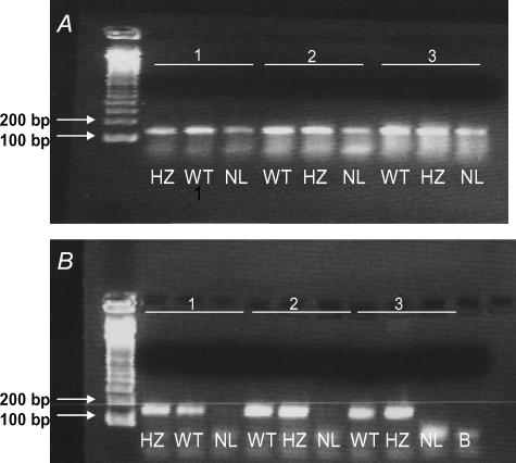
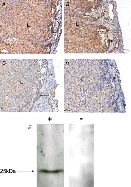
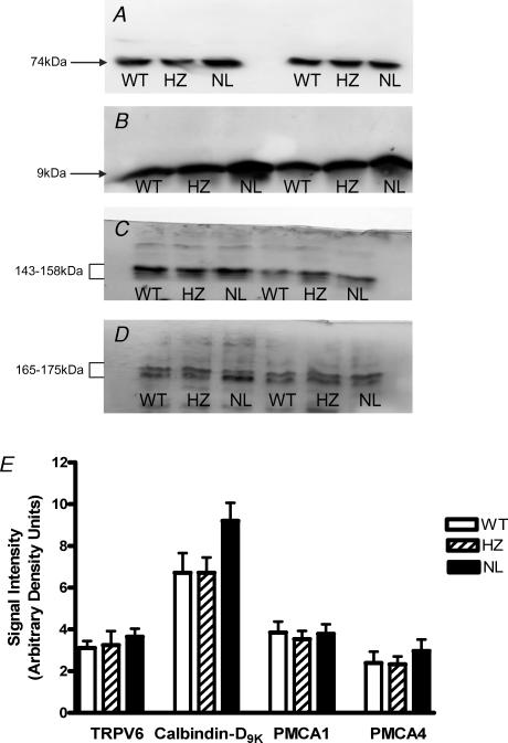
The role of parathyroid hormone-related protein (PTHrP) in fetal calcium homeostasis and placental calcium transport was examined in mice homozygous for the deletion of the PTHrP gene (PTHrP-/- null; NL) compared to PTHrP+/+ (wild-type; WT) and PTHrP+/- (heterozygous; HZ) littermates. Fetal blood ionized calcium was significantly reduced in NL fetuses compared to WT and HZ groups at 18 days of pregnancy (dp) with abolition of the fetomaternal calcium gradient. In situ placental perfusion of the umbilical circulation at 18 dp was used to measure unidirectional clearance of (45)Ca across the placenta in maternofetal ((Ca)K(mf)) and fetoplacental ((Ca)K(fp)) directions; (Ca)K(fp) was < 5% of (Ca)K(mf) for all genotypes. At 18 dp, (Ca)K(mf) across perfused placenta and intact placenta ((Ca)K(mf(intact))) were similar and concordant with net calcium accretion rates in vivo. (Ca)K(mf) was significantly raised in NL fetuses compared to WT and HZ littermates. Calcium accretion was significantly elevated in NL fetuses by 19 dp. Placental calbindin-D(9K) expression in NL fetuses was marginally enhanced (P < 0.07) but expression of TRPV6/ECaC2 and plasma membrane Ca2+-ATPase (PMCA) isoforms 1 and 4 were unaltered. We conclude that PTHrP is an important regulator of fetal calcium homeostasis with its predominant effect being on unidirectional maternofetal transfer, probably mediated by modifying placental calbindin-D(9K) expression. In situ perfusion of mouse placenta is a robust methodology for allowing detailed dissection of placental transfer mechanisms in genetically modified mice.
Figures
References
-
- Abbas SK, Pickard DW, Rodda CP, Heath JH, Hammonds RG, Wood WI, Caple IW, Martin TJ, Care AD. Stimulation of ovine placental calcium transport by purified natural and recombinant parathyroid hormone-related protein (PTHrP) preparations. Q J Exp Physiol. 1989;74:549–552. - PubMed
-
- An B-S, Choi K-C, Lee G-S, Leung PKC, Jeung E-B. Complex regulation of calbindin-D9k in the mouse placenta and extra-embryonic membrane during mid- and late pregnancy. Mol Cell Endocrinol. 2004;214:39–52. - PubMed
-
- Atkinson DE, Boyd RDH, Sibley CP. Placental transfer. In: Neill JD, editor. Knobil and Neill's Physiology of Reproduction. 2. London: Elsevier; 2006. pp. 2787–2846.
-
- Belkacemi L, Bedard I, Simoneau L, Lafond J. Calcium channels, transporters and exchangers in placenta: a review. Cell Calcium. 2005;37:1–8. - PubMed
-
- Bond H, Baker B, Boyd RDH, Cowley E, Glazier JD, Jones CJP, Sibley CP, Ward BS, Husain SM. Artificial perfusion of the fetal circulation of the in situ mouse placenta: methodology and validation. Placenta. 2006a;27(Suppl. A):S69–S75. - PubMed
Publication types
MeSH terms
Substances
Grants and funding
LinkOut - more resources
Full Text Sources
Molecular Biology Databases
Research Materials
Miscellaneous