. 2008 Apr 1;586(7):1963-75.
doi: 10.1113/jphysiol.2007.149815. Epub 2008 Feb 7.

Activity-dependent regulation of tyrosine hydroxylase expression in the enteric nervous system

Affiliations

Activity-dependent regulation of tyrosine hydroxylase expression in the enteric nervous system

J Chevalier et al. J Physiol. .

Abstract

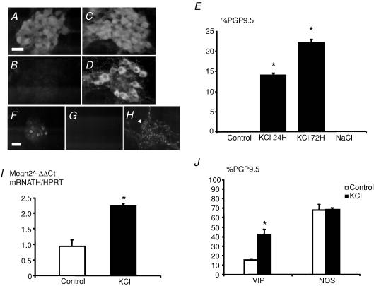
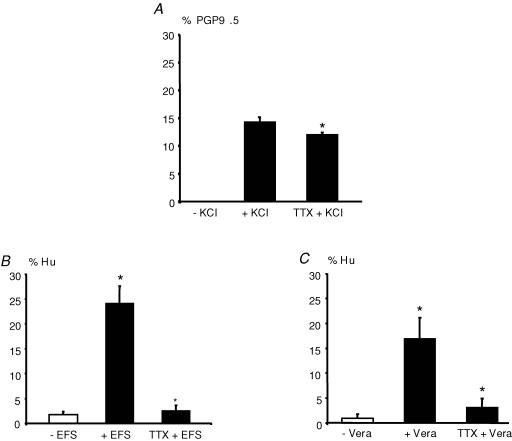
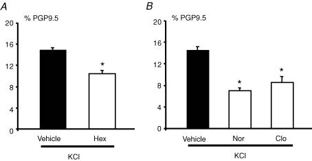
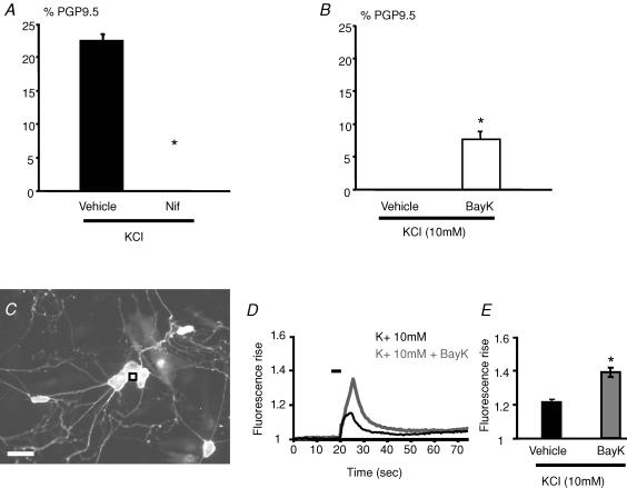
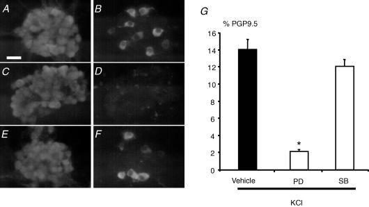
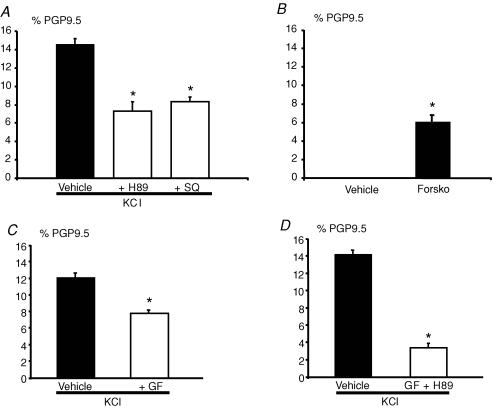
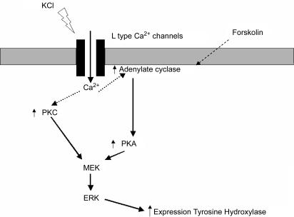
The regulation of neuromediator expression by neuronal activity in the enteric nervous system (ENS) is currently unknown. Using primary cultures of ENS derived from rat embryonic intestine, we have characterized the regulation of tyrosine hydroxylase (TH), a key enzyme involved in the synthesis of dopamine. Depolarization induced either by 40 mm KCl, veratridine or by electrical field stimulation produced a robust and significant increase in the proportion of TH immunoreactive (TH-IR) neurons (total neuronal population was identified with PGP9.5 or Hu) compared to control. This increase in the proportion of TH-IR neurons was significantly reduced by the sodium channel blocker tetrodotoxin (0.5 microm), demonstrating that neuronal activity was critically involved in the effects of these depolarizing stimuli. KCl also increased the proportion of VIP-IR but not nNOS-IR enteric neurons. The KCl-induced increase in TH expression was partly reduced in the presence of the nicotinic receptor antagonist hexamethonium (100 microm), of noradrenaline (1 microm) and of the alpha(2)-adrenoreceptor agonist clonidine (1 microm). Combining pharmacological and calcium imaging studies, we have further shown that L-type calcium channels were involved in the increase of TH expression induced by KCl. Finally, using specific inhibitors, we have shown that both protein kinases A and C as well as the extracellular signal-regulated kinases were required for the increase in the proportion of TH-IR neurons induced by KCl. These results are the first demonstration that TH phenotype of enteric neurons can be regulated by neuronal activity. They could also set the basis for the study of the pathways and mechanisms involved in the neurochemical plasticity observed both during ENS development and in inflammatory enteric neuropathies.


Figures

Figure 1
Figure 1. Characterization of primary culture of ENS
After 14 days in culture, the presence of enteric neurons in primary culture of ENS was assessed by PGP9.5 (A) and Hu immunostaining (B). Enteric glial cells were identified by immunostaining with glial fibrillary acid protein antibodies (C). The neurochemical phenotype was determined by immunostaining using antibodies specific for VIP (D), nNOS (E) and TH (F). Scales bar represents 25 μm.
Figure 2
Figure 2. KCl induces a significant increase in TH and VIP but not nNOS expression in enteric neurons
After 14 days in culture, enteric neurons identified with PGP9.5 (A) were not immunoreactive (IR) for TH (B). Treatment with 40 mm KCl induced a robust increase in TH-IR (D) in PGP9.5-IR enteric neurons (C). The proportion of TH-IR neurons normalized to the total neuronal population identified with PGP9.5 was significantly increased following KCl treatment for 24 and 72 h (E; n = 12; P < 0.001 and n = 8, P < 0.001, respectively, t test). In contrast, incubation of equimolar concentration of NaCl (40 mm) did not modify the proportion of TH-IR neurons (E; n = 8). TH-IR neurons (F) were not dopamine β-hydroxylase (DBH)-IR (G), although the antibody labelled neurons in adult rat ileum (H; arrow). KCl treatment increased TH mRNA transcript expression normalized to HPRT as compared to control (I; n = 8; P < 0.001, t test). Treatment with KCl for 72 h significantly increased the proportion of VIP-IR neurons (J; n = 4; P = 0.003, t test) but not of nNOS-IR neurons (J; n = 4; P = 1, t test). Scale bar represents 25 μm for AD and H and 50 μm for F and G. Data are presented as the mean ±s.e.m.
Figure 3
Figure 3. Neuronal activity is involved in the regulation of TH expression
The increase in the proportion of TH-IR neurons induced by KCl (normalized to the number of PGP9.5-IR neurons) was significantly reduced by 0.5 μm tetrodotoxin (TTX + KCl) (A; n = 5; P = 0.036 one-way ANOVA followed by Turkey's test). EFS (8 h of electrical stimulation) significantly increased the proportion of TH-IR neurons normalized to the number of Hu-IR neurons (+ EFS) as compared to control (− EFS) (B; n = 3; P < 0.001, one-way ANOVA). This effect was blocked following EFS in the presence of 0.5 μm tetrodotoxin (TTX + EFS) (B). Treatment of primary culture of enteric neurons with 30 μm veratridine (24 h) (+ Vera) significantly increased the proportion of TH-IR neurons (normalized to the number of Hu-IR neurons) as compared to control (− Vera) (C; n = 4; P = 0.022, one-way ANOVA). These effects were also blocked in the presence of 0.5 μm tetrodotoxin (TTX + Vera) (C).
Figure 4
Figure 4. Cholinergic and noradrenergic pathways regulate the KCl-induced increase in the proportion of TH-immunoreactive neurons
The increase in the proportion of TH-IR neurons induced by KCl was significantly reduced by hexamethonium (Hex, 100 μm) as compared to control (Vehicle) (A; n = 9; P < 0.001, t test). The KCl-induced increase in the proportion of TH-IR neurons was also significantly reduced by noradrenaline (Nor, 1 μm) and clonidine (Clo, 1 μm) as compared to control (Vehicle) (B; n = 4; P≤ 0.001 and n = 4; P = 0.005, respectively, t test). Data are presented as the mean ±s.e.m.
Figure 5
Figure 5. L-type calcium channels are involved in the KCl-induced increase in the proportion of TH-immunoreactive neurons
The increase in the proportion of TH-IR neurons induced by KCl was completely prevented by nifedipine (Nif, 1 μm) (A; n = 6, P = 0.002, Mann–Whitney rank sum test). Treatment with BayK-8644 (BayK, 1 μm) in the presence of 10 mm KCl significantly increased the proportion of TH-IR neurons as compared to control (Vehicle) (B; n = 6, P = 0.002, Mann–Whitney rank sum test). Intracellular calcium measurements were performed on cells loaded with Fluo-4 AM and identification of neurons was performed by application of 75 mm KCl (C) and Ca2+ signals were calculated from individual neurons (e.g. as indicated with a black square). Microejection of 10 mm KCl (5 s ejection duration) onto neurons induced a transient relative fluorescence rise (F1/F0) of the Ca2+ indicator Fluo-4 over time (D). Microejection of 10 mm KCl in the presence of BayK-8644 (dark grey line, 1 μm) induced a larger transient fluorescence rise than with 10 mm KCl (D). The traces in this example are from the neuron marked (black square) in C. The amplitude of the transient relative fluorescence rise induced by 10 mm KCl is significantly larger in the presence of BayK-8644 (E; P < 0.0001; n = 93 neurons). Scale bar represents 50 μm. Data are presented as the mean ±s.e.m.
Figure 6
Figure 6. ERK- but not p38-dependent pathways mediate the KCl-induced increase in the proportion of TH-immunoreactive neurons
Photomicrographs illustrate that in the presence of KCl a proportion of PGP9.5-IR neurons (A) was TH-IR (B). In the presence of KCl and the inhibitor of ERK pathways (PD98059; 50 μm), no PGP9.5-IR neurons (C) were TH-IR (D). In contrast, in the presence of KCl and the inhibitor of p38 pathways (SB203580; 10 μm) a proportion of PGP9.5-IR neurons (E) was still TH-IR (F). Quantification of the results demonstrated that PD98059 (PD), but not SB203580 (SB) significantly reduced the KCl-induced increase in the proportion of TH-IR neurons (G; P < 0.001, n = 5,t test). Scale bar represents 25 μm. Data are presented as the mean ±s.e.m.
Figure 7
Figure 7. Both, cAMP- and PKC-dependent pathways mediate the KCl-induced increase in TH-immunoreactive neurons
The increase in the proportion of TH-IR neurons induced by KCl was significantly reduced by an inhibitor of adenylate cyclase, SQ22536 (SQ, 100 μm; n = 5; P = 0.008, t test), and by an inhibitor of PKA, H89 (A) (H89 2 μm; n = 5; P≤ 0.001, t test). Forskolin, an activator of adenylate cyclase, induced a significant increase in the proportion of TH-IR neurons as compared to control (B) (Forsko, 20 μm; n = 5; P = 0.001, t test). The increase in the proportion of TH-IR neurons induced by KCl was also significantly reduced by the inhibitor of PKC, GF109203X (C) (GF 1 μm; n = 4; P = 0.029, t test). In the presence of both PKC and PKA inhibitors, the increase in the proportion of TH-IR neurons induced by KCl was further significantly reduced as compared to the condition with the inhibitors alone (D) (n = 5; P≤ 0.001, t test). Data are presented as the mean ±s.e.m.
Figure 8
Figure 8. Signalling pathways involved in the activity-dependent regulation of TH expression in enteric neurons
Neuronal activation by depolarization results in opening of L-type calcium channels leading to an increase in calcium entry. This increase in intracellular calcium induces an activation of ERKs through an activation of both PKA and PKC, resulting in an increase in TH expression.

References

    1. Alessi DR, Cuenda A, Cohen P, Dudley DT, Saltiel AR. PD 098059 is a specific inhibitor of the activation of mitogen-activated protein kinase kinase in vitro and in vivo. J Biol Chem. 1995;270:27489–27494. - PubMed
    1. Anlauf M, Schafer MK, Eiden L, Weihe E. Chemical coding of the human gastrointestinal nervous system: cholinergic, VIPergic, and catecholaminergic phenotypes. J Comp Neurol. 2003;459:90–111. - PubMed
    1. Baldassa S, Zippel R, Sturani E. Depolarization- induced signaling to Ras, Rap1 and MAPKs in cortical neurons. Brain Res Mol Brain Res. 2003;119:111–122. - PubMed
    1. Belai A, Boulos PB, Robson T, Burnstock G. Neurochemical coding in the small intestine of patients with Crohn's disease. Gut. 1997;40:767–774. - PMC - PubMed
    1. Brosenitsch TA, Katz DM. Physiological patterns of electrical stimulation can induce neuronal gene expression by activating N-type calcium channels. J Neurosci. 2001;21:2571–2579. - PMC - PubMed

Publication types

MeSH terms

LinkOut - more resources