

3D rotational angiography: the new gold standard in the detection of additional intracranial aneurysms
- PMID: 18258703
- PMCID: PMC8128578
- DOI: 10.3174/ajnr.A0964
3D rotational angiography: the new gold standard in the detection of additional intracranial aneurysms
Abstract
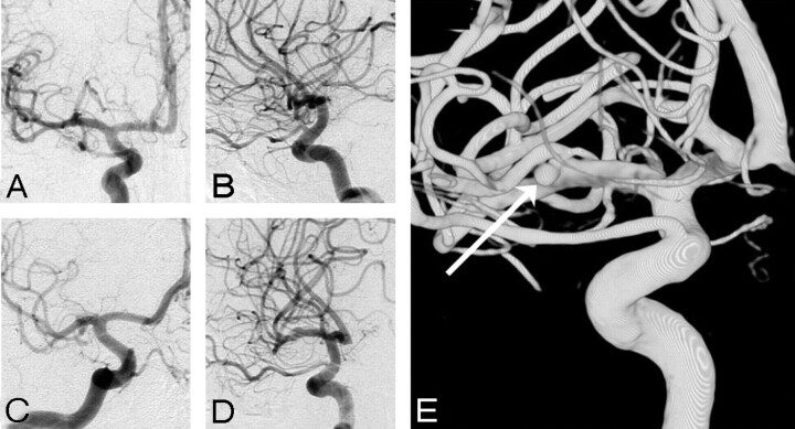
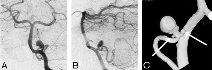
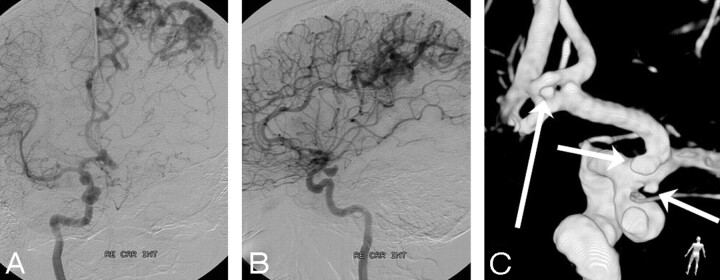
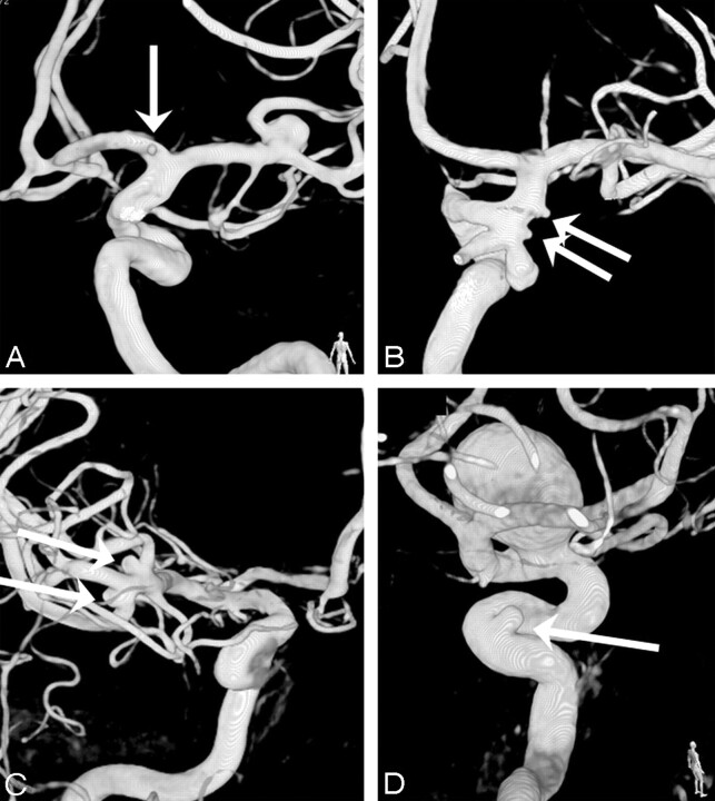
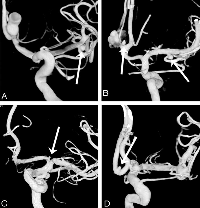
Background and purpose: During surgery of symptomatic aneurysms, additional small angiographic occult aneurysms are commonly found. With 3D rotational angiography (3DRA) small aneurysms are more easily depicted than with digital subtraction angiography (DSA). In this study we compare 3DRA with DSA in the depiction of small additional aneurysms.
Materials and methods: Three hundred fifty 3D datasets of 1 vascular tree of 350 patients with at least 1 intracranial aneurysm on the dataset were re-evaluated for the presence of additional aneurysms by 2 observers in consensus. Two other observers, blinded to the 3D images, re-evaluated DSA images of the same 350 vascular trees for these additional aneurysms. Results were compared.
Results: In 350 3D datasets, 350 target aneurysms and 94 additional aneurysms were detected. The mean size of 94 additional aneurysms was 3.54 mm (median, 3; range, 0.5-17 mm). The proportion of aneurysms <or=3 mm was significantly higher in additional aneurysms (61 of 94, 65%) than in the target aneurysms (61 of 350, 17%) (chi(2), P < .0001). Of 94 additional aneurysms, 27 (29%) were missed on DSA by both observers. The mean size of the missed aneurysms was 1.94 mm (median, 2; range, 0.5-4 mm). The proportion of aneurysms <or=3 mm in missed additional aneurysms (26 of 27, 96%) was significantly higher than that in all additional aneurysms (61 of 94, 65%) (chi(2), P = .0035). The location of missed additional aneurysms was not different from the location of all additional aneurysms.
Conclusion: 3DRA depicts considerably more small (<or=3 mm) additional aneurysms than DSA. In selected patients, accurate detection of these aneurysms may have consequences for the choice of treatment technique and for the frequency and duration of imaging follow-up.
Figures
References
-
- Kouskouras C, Charitanti A, Giavroglou C, et al. Intracranial aneurysms: evaluation using CTA and MRA—correlation with DSA and intraoperative findings. Neuroradiology 2004;46:842–50 - PubMed
-
- Jayaraman MV, Mayo-Smith WW, Tung GA, et al. Detection of intracranial aneurysms: multi-detector row CT angiography compared with DSA. Radiology 2004;230:510–18 - PubMed
-
- Inamasu J, Suga S, Horiguchi T, et al. Cerebral micro aneurysms found incidentally during aneurysm surgery. Neurol Res 2001;23:304–08 - PubMed
-
- Karasawa H, Matsumoto H, Naito H, et al. Angiographically unrecognized microaneurysms: intraoperative observation and operative technique. Acta Neurochir (Wien) 1997;139:416–19, discussion 419–20 - PubMed
Publication types
MeSH terms
LinkOut - more resources
Full Text Sources
Medical